- PDB-3aur: Crystal structure of the human vitamin D receptor ligand binding ... -
+
Open data
ID or keywords:
Loading...
-
Basic information
Entry
Database: PDB / ID: 3aur
Title
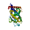
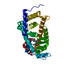
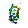
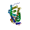
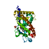
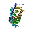
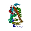
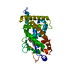
Crystal structure of the human vitamin D receptor ligand binding domain complexed with Yne-diene type analog of active 14-epi-2beta-methyl-19-norvitamin D3
Components
Vitamin D3 receptor
Keywords
TRANSCRIPTION / HORMONE RECEPTOR
Function / homology
Function and homology information
apoptotic process involved in mammary gland involution / response to bile acid / Vitamin D (calciferol) metabolism / positive regulation of apoptotic process involved in mammary gland involution / calcitriol binding / lithocholic acid binding / nuclear receptor-mediated bile acid signaling pathway / bile acid nuclear receptor activity / positive regulation of keratinocyte differentiation / vitamin D binding ...apoptotic process involved in mammary gland involution / response to bile acid / Vitamin D (calciferol) metabolism / positive regulation of apoptotic process involved in mammary gland involution / calcitriol binding / lithocholic acid binding / nuclear receptor-mediated bile acid signaling pathway / bile acid nuclear receptor activity / positive regulation of keratinocyte differentiation / vitamin D binding / vitamin D receptor signaling pathway / positive regulation of vitamin D receptor signaling pathway / phosphate ion transmembrane transport / intestinal absorption / mammary gland branching involved in pregnancy / decidualization / negative regulation of keratinocyte proliferation / positive regulation of bone mineralization / nuclear retinoid X receptor binding / retinoic acid receptor signaling pathway / intracellular receptor signaling pathway / lactation / SUMOylation of intracellular receptors / skeletal system development / Nuclear Receptor transcription pathway / mRNA transcription by RNA polymerase II / nuclear receptor activity / RNA polymerase II transcription regulator complex / cell morphogenesis / intracellular calcium ion homeostasis / calcium ion transport / DNA-binding transcription factor activity, RNA polymerase II-specific / cell differentiation / signaling receptor complex / RNA polymerase II cis-regulatory region sequence-specific DNA binding / negative regulation of cell population proliferation / negative regulation of DNA-templated transcription / positive regulation of gene expression / chromatin / negative regulation of transcription by RNA polymerase II / positive regulation of transcription by RNA polymerase II / DNA binding / zinc ion binding / nucleoplasm / nucleus / cytosol Similarity search - Function
Mass: 18.015 Da / Num. of mol.: 75 / Source method: isolated from a natural source / Formula: H2O
Nonpolymer details
THE LIGAND IN THIS ENTRY HAS INDEED A DIFFERENT STEREOCHEMISTRY OF ATOM C26 FROM CA9 STRUCTURE. BUT ...THE LIGAND IN THIS ENTRY HAS INDEED A DIFFERENT STEREOCHEMISTRY OF ATOM C26 FROM CA9 STRUCTURE. BUT THIS COMPOUND TURN OVER IN THIS STRUCTURE.
Sequence details
THIS ENTRY IS DELETION MUTANT. RESIDUES 165-215 WERE DELETED.
-
Experimental details
-
Experiment
Experiment
Method: X-RAY DIFFRACTION / Number of used crystals: 1
-
Sample preparation
Crystal
Density Matthews: 2.52 Å3/Da / Density % sol: 51.22 %
Crystal grow
Temperature: 293 K / Method: vapor diffusion, hanging drop / pH: 6.5 Details: 0.1M MES, 1.2-1.6M ammonium sulfate , pH 6.5, VAPOR DIFFUSION, HANGING DROP, temperature 293K
Resolution: 2.21→36.77 Å / Cor.coef. Fo:Fc: 0.939 / Cor.coef. Fo:Fc free: 0.899 / SU B: 5.33 / SU ML: 0.138 / Cross valid method: THROUGHOUT / ESU R: 0.259 / ESU R Free: 0.222 / Stereochemistry target values: MAXIMUM LIKELIHOOD / Details: HYDROGENS HAVE BEEN ADDED IN THE RIDING POSITIONS
Rfactor
Num. reflection
% reflection
Selection details
Rfree
0.25427
761
5 %
RANDOM
Rwork
0.19073
-
-
-
obs
0.19393
14435
96.96 %
-
Solvent computation
Ion probe radii: 0.8 Å / Shrinkage radii: 0.8 Å / VDW probe radii: 1.2 Å / Solvent model: MASK
Displacement parameters
Biso mean: 34.598 Å2
Baniso -1
Baniso -2
Baniso -3
1-
0 Å2
0 Å2
0 Å2
2-
-
0.01 Å2
0 Å2
3-
-
-
0 Å2
Refinement step
Cycle: LAST / Resolution: 2.21→36.77 Å
Protein
Nucleic acid
Ligand
Solvent
Total
Num. atoms
2018
0
30
75
2123
Refine LS restraints
Refine-ID
Type
Dev ideal
Dev ideal target
Number
X-RAY DIFFRACTION
r_bond_refined_d
0.022
0.022
2090
X-RAY DIFFRACTION
r_bond_other_d
X-RAY DIFFRACTION
r_angle_refined_deg
1.977
2.003
2832
X-RAY DIFFRACTION
r_angle_other_deg
X-RAY DIFFRACTION
r_dihedral_angle_1_deg
6.818
5
251
X-RAY DIFFRACTION
r_dihedral_angle_2_deg
34.62
24.286
91
X-RAY DIFFRACTION
r_dihedral_angle_3_deg
17.987
15
383
X-RAY DIFFRACTION
r_dihedral_angle_4_deg
17.555
15
13
X-RAY DIFFRACTION
r_chiral_restr
0.13
0.2
328
X-RAY DIFFRACTION
r_gen_planes_refined
0.009
0.02
1529
X-RAY DIFFRACTION
r_gen_planes_other
X-RAY DIFFRACTION
r_nbd_refined
0.226
0.2
1037
X-RAY DIFFRACTION
r_nbd_other
X-RAY DIFFRACTION
r_nbtor_refined
0.309
0.2
1447
X-RAY DIFFRACTION
r_nbtor_other
X-RAY DIFFRACTION
r_xyhbond_nbd_refined
0.169
0.2
121
X-RAY DIFFRACTION
r_xyhbond_nbd_other
X-RAY DIFFRACTION
r_metal_ion_refined
X-RAY DIFFRACTION
r_metal_ion_other
X-RAY DIFFRACTION
r_symmetry_vdw_refined
0.261
0.2
51
X-RAY DIFFRACTION
r_symmetry_vdw_other
X-RAY DIFFRACTION
r_symmetry_hbond_refined
0.11
0.2
6
X-RAY DIFFRACTION
r_symmetry_hbond_other
X-RAY DIFFRACTION
r_symmetry_metal_ion_refined
X-RAY DIFFRACTION
r_symmetry_metal_ion_other
X-RAY DIFFRACTION
r_mcbond_it
1.34
1.5
1323
X-RAY DIFFRACTION
r_mcbond_other
X-RAY DIFFRACTION
r_mcangle_it
2.074
2
2065
X-RAY DIFFRACTION
r_scbond_it
3.124
3
868
X-RAY DIFFRACTION
r_scangle_it
4.66
4.5
767
X-RAY DIFFRACTION
r_rigid_bond_restr
X-RAY DIFFRACTION
r_sphericity_free
X-RAY DIFFRACTION
r_sphericity_bonded
LS refinement shell
Resolution: 2.213→2.271 Å / Total num. of bins used: 20
Rfactor
Num. reflection
% reflection
Rfree
0.322
60
-
Rwork
0.181
1021
-
obs
-
-
96.77 %
+
About Yorodumi
-
News
-
Feb 9, 2022. New format data for meta-information of EMDB entries
New format data for meta-information of EMDB entries
Version 3 of the EMDB header file is now the official format.
The previous official version 1.9 will be removed from the archive.
In the structure databanks used in Yorodumi, some data are registered as the other names, "COVID-19 virus" and "2019-nCoV". Here are the details of the virus and the list of structure data.
Jan 31, 2019. EMDB accession codes are about to change! (news from PDBe EMDB page)
EMDB accession codes are about to change! (news from PDBe EMDB page)
The allocation of 4 digits for EMDB accession codes will soon come to an end. Whilst these codes will remain in use, new EMDB accession codes will include an additional digit and will expand incrementally as the available range of codes is exhausted. The current 4-digit format prefixed with “EMD-” (i.e. EMD-XXXX) will advance to a 5-digit format (i.e. EMD-XXXXX), and so on. It is currently estimated that the 4-digit codes will be depleted around Spring 2019, at which point the 5-digit format will come into force.
The EM Navigator/Yorodumi systems omit the EMD- prefix.
Related info.:Q: What is EMD? / ID/Accession-code notation in Yorodumi/EM Navigator
Yorodumi is a browser for structure data from EMDB, PDB, SASBDB, etc.
This page is also the successor to EM Navigator detail page, and also detail information page/front-end page for Omokage search.
The word "yorodu" (or yorozu) is an old Japanese word meaning "ten thousand". "mi" (miru) is to see.
Related info.:EMDB / PDB / SASBDB / Comparison of 3 databanks / Yorodumi Search / Aug 31, 2016. New EM Navigator & Yorodumi / Yorodumi Papers / Jmol/JSmol / Function and homology information / Changes in new EM Navigator and Yorodumi
 Movie
Movie Controller

 Yorodumi
 Open data
 Basic information
 Components
 Keywords
 Function and homology information
 Homo sapiens (human)
 X-RAY DIFFRACTION /
  Authors
 Citation
 Structure visualization
 Downloads & links
 Other downloads
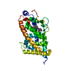


 PDBj
PDBj

 Assembly


 Mass: 414.621 Da / Num. of mol.: 1 / Source method: obtained synthetically / Formula: C27H42O3
 Mass: 18.015 Da / Num. of mol.: 75 / Source method: isolated from a natural source / Formula: H2O
 Sample preparation
 / Beamline: BL41XU / Wavelength: 1 Å
 Processing