[English] 日本語
Yorodumi
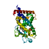
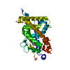
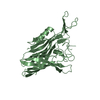
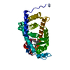
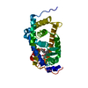
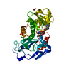
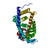
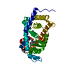
- PDB-5ysy: Crystal structure of the human vitamin D receptor ligand binding ... -

+
Open data


ID or keywords:

Loading...

-
Basic information

Entry
Database: PDB / ID: 5ysy
TitleCrystal structure of the human vitamin D receptor ligand binding domain complexed with (1R,2R,3R)-5-[(E)-2-{(1R,3aS,7aR)-1-[(R)-6-hydroxy-6-methylheptan-2-yl]-7a-methyl-2,3,3a,6,7,7a-hexahydro-1H-inden-4-yl}vinyl]-2-(3-hydroxypropyl)cyclohex-4-ene-1,3-diol
ComponentsVitamin D3 receptor
KeywordsTRANSCRIPTION / Active ligand / Complex
Function / homology
Function and homology information


regulation of calcidiol 1-monooxygenase activity / positive regulation of vitamin D 24-hydroxylase activity / bile acid nuclear receptor activity / response to bile acid / Vitamin D (calciferol) metabolism / phosphate ion transmembrane transport / apoptotic process involved in mammary gland involution / calcitriol binding / positive regulation of apoptotic process involved in mammary gland involution / lithocholic acid binding ...regulation of calcidiol 1-monooxygenase activity / positive regulation of vitamin D 24-hydroxylase activity / bile acid nuclear receptor activity / response to bile acid / Vitamin D (calciferol) metabolism / phosphate ion transmembrane transport / apoptotic process involved in mammary gland involution / calcitriol binding / positive regulation of apoptotic process involved in mammary gland involution / lithocholic acid binding / positive regulation of keratinocyte differentiation / positive regulation of vitamin D receptor signaling pathway / vitamin D receptor signaling pathway / bile acid signaling pathway / intestinal absorption / mammary gland branching involved in pregnancy / decidualization / negative regulation of keratinocyte proliferation / positive regulation of bone mineralization / nuclear retinoid X receptor binding / lactation / skeletal system development / SUMOylation of intracellular receptors / mRNA transcription by RNA polymerase II / cell morphogenesis / intracellular calcium ion homeostasis / Nuclear Receptor transcription pathway / RNA polymerase II transcription regulator complex / nuclear receptor activity / calcium ion transport / cell differentiation / receptor complex / DNA-binding transcription factor activity, RNA polymerase II-specific / RNA polymerase II cis-regulatory region sequence-specific DNA binding / negative regulation of cell population proliferation / negative regulation of DNA-templated transcription / chromatin / positive regulation of gene expression / negative regulation of transcription by RNA polymerase II / positive regulation of transcription by RNA polymerase II / DNA binding / zinc ion binding / nucleoplasm / nucleus / cytosol
Similarity search - Function
Vitamin D receptor / VDR, DNA-binding domain / Retinoid X Receptor / Retinoid X Receptor / Nuclear hormone receptor / Nuclear hormones receptors DNA-binding region signature. / Zinc finger, nuclear hormone receptor-type / Zinc finger, C4 type (two domains) / Nuclear hormone receptors DNA-binding domain profile. / c4 zinc finger in nuclear hormone receptors ...Vitamin D receptor / VDR, DNA-binding domain / Retinoid X Receptor / Retinoid X Receptor / Nuclear hormone receptor / Nuclear hormones receptors DNA-binding region signature. / Zinc finger, nuclear hormone receptor-type / Zinc finger, C4 type (two domains) / Nuclear hormone receptors DNA-binding domain profile. / c4 zinc finger in nuclear hormone receptors / Nuclear hormone receptor, ligand-binding domain / Nuclear hormone receptor-like domain superfamily / Ligand-binding domain of nuclear hormone receptor / Nuclear receptor (NR) ligand-binding (LBD) domain profile. / Ligand binding domain of hormone receptors / Zinc finger, NHR/GATA-type / Orthogonal Bundle / Mainly Alpha
Similarity search - Domain/homology
Chem-90L / Vitamin D3 receptor
Similarity search - Component
Biological speciesHomo sapiens (human)
MethodX-RAY DIFFRACTION / SYNCHROTRON / MOLECULAR REPLACEMENT / Resolution: 2 Å
AuthorsTakimoto-Kamimura, M. / Kakuda, S.
CitationJournal: Org. Biomol. Chem. / Year: 2018
Title: Effects of 2-substitution on 14-epi-19-nortachysterol-mediated biological events: based on synthesis and X-ray co-crystallographic analysis with the human vitamin D receptor.
Authors: Sawada, D. / Kakuda, S. / Takeuchi, A. / Kawagoe, F. / Takimoto-Kamimura, M. / Kittaka, A.
History
DepositionNov 16, 2017Deposition site: PDBJ / Processing site: PDBJ
Revision 1.0Apr 11, 2018Provider: repository / Type: Initial release
Revision 1.1Apr 18, 2018Group: Data collection / Database references / Category: citation
Item: _citation.journal_volume / _citation.page_first / _citation.page_last
Revision 1.2Nov 22, 2023Group: Data collection / Database references ...Data collection / Database references / Derived calculations / Refinement description / Structure summary
Category: chem_comp / chem_comp_atom ...chem_comp / chem_comp_atom / chem_comp_bond / database_2 / entity / pdbx_entity_nonpoly / pdbx_initial_refinement_model
Item: _chem_comp.name / _chem_comp.pdbx_synonyms ..._chem_comp.name / _chem_comp.pdbx_synonyms / _database_2.pdbx_DOI / _database_2.pdbx_database_accession / _entity.pdbx_description / _pdbx_entity_nonpoly.name

-
Structure visualization

Structure viewerMolecule:
MolmilJmol/JSmol

Downloads & links

-
Assembly

Deposited unit
A: Vitamin D3 receptor
hetero molecules


Theoretical massNumber of molelcules
Total (without water)29,4092
Polymers28,9481
Non-polymers4611
Water3,153175
1


  • Idetical with deposited unit
  • defined by author&software
TypeNameSymmetry operationNumber
identity operation1_555x,y,z1
Buried area0 Å2
ΔGint0 kcal/mol
Surface area11980 Å2
MethodPISA
Unit cell
Length a, b, c (Å)44.682, 51.538, 132.651
Angle α, β, γ (deg.)90.00, 90.00, 90.00
Int Tables number19
Space group name H-MP212121

-
Components

#1: Protein Vitamin D3 receptor / VDR / 1 / 25-dihydroxyvitamin D3 receptor / Nuclear receptor subfamily 1 group I member 1


Mass: 28948.420 Da / Num. of mol.: 1 / Fragment: ligand binding domain
Source method: isolated from a genetically manipulated source
Source: (gene. exp.) Homo sapiens (human) / Gene: VDR, NR1I1 / Production host: Escherichia coli (E. coli) / References: UniProt: P11473
#2: Chemical ChemComp-90L / (1R,2R,3R)-5-[(E)-2-[(1R,3aS,7aR)-7a-methyl-1-[(2R)-6-methyl-6-oxidanyl-heptan-2-yl]-1,2,3,3a,6,7-hexahydroinden-4-yl]e thenyl]-2-(3-oxidanylpropyl)cyclohex-4-ene-1,3-diol / (1R,2R,3R)-5-[(E)-2-{(1R,3aS,7aR)-1-[(R)-6-hydroxy-6-methylheptan-2-yl]-7a-methyl-2,3,3a,6,7,7a-hexahydro-1H-inden-4-yl }vinyl]-2-(3-hydroxypropyl)cyclohex-4-ene-1,3-diol


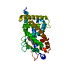
Mass: 460.689 Da / Num. of mol.: 1 / Source method: obtained synthetically / Formula: C29H48O4 / Feature type: SUBJECT OF INVESTIGATION
#3: Water ChemComp-HOH / water / Water


Mass: 18.015 Da / Num. of mol.: 175 / Source method: isolated from a natural source / Formula: H2O

-
Experimental details

-
Experiment

ExperimentMethod: X-RAY DIFFRACTION / Number of used crystals: 1

-
Sample preparation

CrystalDensity Matthews: 2.64 Å3/Da / Density % sol: 53.37 %
Crystal growTemperature: 277 K / Method: vapor diffusion, hanging drop / pH: 6.5 / Details: 50mM Mes, 1.4M ammonium sulphate

-
Data collection

DiffractionMean temperature: 100 K
Diffraction sourceSource: SYNCHROTRON / Site: Photon Factory / Beamline: AR-NE3A / Wavelength: 1 Å
DetectorType: ADSC QUANTUM 315r / Detector: CCD / Date: Jun 20, 2011
RadiationProtocol: SINGLE WAVELENGTH / Monochromatic (M) / Laue (L): M / Scattering type: x-ray
Radiation wavelengthWavelength: 1 Å / Relative weight: 1
ReflectionResolution: 2→50 Å / Num. obs: 21471 / % possible obs: 99.9 % / Redundancy: 7.7 % / Rmerge(I) obs: 0.096 / Net I/σ(I): 35.3
Reflection shellRmerge(I) obs: 0.431

-
Processing

Software
NameVersionClassification
REFMAC5.2.0019refinement
HKL-2000data reduction
SCALA3.3.20data scaling
MOLREP11.0.05phasing
RefinementMethod to determine structure: MOLECULAR REPLACEMENT
Starting model: 1DB1
Resolution: 2→50 Å / Cor.coef. Fo:Fc: 0.946 / Cor.coef. Fo:Fc free: 0.913 / SU B: 3.58 / SU ML: 0.103 / Cross valid method: THROUGHOUT / ESU R: 0.174 / ESU R Free: 0.165
Details: 1. The authors state they removed the long loop 165-215 for the crystallization with direct lincage with Gly 164 and Ser 216, so this region recieved quite large steric hindrance and resuled ...Details: 1. The authors state they removed the long loop 165-215 for the crystallization with direct lincage with Gly 164 and Ser 216, so this region recieved quite large steric hindrance and resuled abnormal bond length as usual, and that the bond is existing in real but the bond distance is abnormal by the disorder by the steric hindrances. 2. HYDROGENS HAVE BEEN ADDED IN THE RIDING POSITIONS
RfactorNum. reflection% reflectionSelection details
Rfree0.24223 1097 5.1 %RANDOM
Rwork0.18936 ---
obs0.19196 20302 99.93 %-
Solvent computationIon probe radii: 0.8 Å / Shrinkage radii: 0.8 Å / VDW probe radii: 1.2 Å
Displacement parametersBiso mean: 24.206 Å2
Baniso -1Baniso -2Baniso -3
1--0.16 Å20 Å20 Å2
2---0.34 Å20 Å2
3---0.5 Å2
Refinement stepCycle: 1 / Resolution: 2→50 Å
ProteinNucleic acidLigandSolventTotal
Num. atoms2027 0 33 175 2235
Refine LS restraints
Refine-IDTypeDev idealDev ideal targetNumber
X-RAY DIFFRACTIONr_bond_refined_d0.0180.0222147
X-RAY DIFFRACTIONr_bond_other_d
X-RAY DIFFRACTIONr_angle_refined_deg1.7292.0022914
X-RAY DIFFRACTIONr_angle_other_deg
X-RAY DIFFRACTIONr_dihedral_angle_1_deg7.0565263
X-RAY DIFFRACTIONr_dihedral_angle_2_deg35.81524.25594
X-RAY DIFFRACTIONr_dihedral_angle_3_deg14.93615392
X-RAY DIFFRACTIONr_dihedral_angle_4_deg18.6891514
X-RAY DIFFRACTIONr_chiral_restr0.1130.2333
X-RAY DIFFRACTIONr_gen_planes_refined0.0070.021594
X-RAY DIFFRACTIONr_gen_planes_other
X-RAY DIFFRACTIONr_nbd_refined0.2270.21010
X-RAY DIFFRACTIONr_nbd_other
X-RAY DIFFRACTIONr_nbtor_refined0.3040.21511
X-RAY DIFFRACTIONr_nbtor_other
X-RAY DIFFRACTIONr_xyhbond_nbd_refined0.1540.2135
X-RAY DIFFRACTIONr_xyhbond_nbd_other
X-RAY DIFFRACTIONr_metal_ion_refined
X-RAY DIFFRACTIONr_metal_ion_other
X-RAY DIFFRACTIONr_symmetry_vdw_refined0.2240.248
X-RAY DIFFRACTIONr_symmetry_vdw_other
X-RAY DIFFRACTIONr_symmetry_hbond_refined0.1740.214
X-RAY DIFFRACTIONr_symmetry_hbond_other
X-RAY DIFFRACTIONr_symmetry_metal_ion_refined
X-RAY DIFFRACTIONr_symmetry_metal_ion_other
X-RAY DIFFRACTIONr_mcbond_it1.161.51364
X-RAY DIFFRACTIONr_mcbond_other
X-RAY DIFFRACTIONr_mcangle_it1.86922130
X-RAY DIFFRACTIONr_mcangle_other
X-RAY DIFFRACTIONr_scbond_it2.4493886
X-RAY DIFFRACTIONr_scbond_other
X-RAY DIFFRACTIONr_scangle_it3.7024.5784
X-RAY DIFFRACTIONr_scangle_other
X-RAY DIFFRACTIONr_long_range_B_refined
X-RAY DIFFRACTIONr_long_range_B_other
X-RAY DIFFRACTIONr_rigid_bond_restr
X-RAY DIFFRACTIONr_sphericity_free
X-RAY DIFFRACTIONr_sphericity_bonded
LS refinement shellResolution: 2.001→2.053 Å / Total num. of bins used: 20
RfactorNum. reflection% reflection
Rfree0.273 81 -
Rwork0.193 1463 -
obs--99.94 %

+
About Yorodumi

-
News

-
Feb 9, 2022. New format data for meta-information of EMDB entries

New format data for meta-information of EMDB entries

  • Version 3 of the EMDB header file is now the official format.
  • The previous official version 1.9 will be removed from the archive.

Related info.:EMDB header

External links:wwPDB to switch to version 3 of the EMDB data model

-
Aug 12, 2020. Covid-19 info

Covid-19 info

URL: https://pdbj.org/emnavi/covid19.php

New page: Covid-19 featured information page in EM Navigator.

Related info.:Covid-19 info / Mar 5, 2020. Novel coronavirus structure data

+
Mar 5, 2020. Novel coronavirus structure data

Novel coronavirus structure data

Related info.:Yorodumi Speices / Aug 12, 2020. Covid-19 info

External links:COVID-19 featured content - PDBj / Molecule of the Month (242):Coronavirus Proteases

+
Jan 31, 2019. EMDB accession codes are about to change! (news from PDBe EMDB page)

EMDB accession codes are about to change! (news from PDBe EMDB page)

  • The allocation of 4 digits for EMDB accession codes will soon come to an end. Whilst these codes will remain in use, new EMDB accession codes will include an additional digit and will expand incrementally as the available range of codes is exhausted. The current 4-digit format prefixed with “EMD-” (i.e. EMD-XXXX) will advance to a 5-digit format (i.e. EMD-XXXXX), and so on. It is currently estimated that the 4-digit codes will be depleted around Spring 2019, at which point the 5-digit format will come into force.
  • The EM Navigator/Yorodumi systems omit the EMD- prefix.

Related info.:Q: What is EMD? / ID/Accession-code notation in Yorodumi/EM Navigator

External links:EMDB Accession Codes are Changing Soon! / Contact to PDBj

+
Jul 12, 2017. Major update of PDB

Major update of PDB

  • wwPDB released updated PDB data conforming to the new PDBx/mmCIF dictionary.
  • This is a major update changing the version number from 4 to 5, and with Remediation, in which all the entries are updated.
  • In this update, many items about electron microscopy experimental information are reorganized (e.g. em_software).
  • Now, EM Navigator and Yorodumi are based on the updated data.

External links:wwPDB Remediation / Enriched Model Files Conforming to OneDep Data Standards Now Available in the PDB FTP Archive

-
Yorodumi

Thousand views of thousand structures

  • Yorodumi is a browser for structure data from EMDB, PDB, SASBDB, etc.
  • This page is also the successor to EM Navigator detail page, and also detail information page/front-end page for Omokage search.
  • The word "yorodu" (or yorozu) is an old Japanese word meaning "ten thousand". "mi" (miru) is to see.

Related info.:EMDB / PDB / SASBDB / Comparison of 3 databanks / Yorodumi Search / Aug 31, 2016. New EM Navigator & Yorodumi / Yorodumi Papers / Jmol/JSmol / Function and homology information / Changes in new EM Navigator and Yorodumi

Read more