 Movie
Movie Controller


+ Open data

- Basic information
| Entry | Database: PDB / ID: 3asi | ||||||
|---|---|---|---|---|---|---|---|
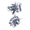
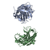
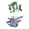
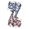
| Title | Alpha-Neurexin-1 ectodomain fragment; LNS5-EGF3-LNS6 | ||||||
 Components Components | Neurexin-1-alpha | ||||||
 Keywords Keywords | CELL ADHESION / Beta-sandwich / synapse maturation / Neuroligin / N-glycosylation / Membrane | ||||||
| Function / homology |  Function and homology information Function and homology informationneuroligin family protein binding / presynaptic membrane / cell adhesion / metal ion binding Similarity search - Function | ||||||
| Biological species |  | ||||||
| Method |  X-RAY DIFFRACTION / SYNCHROTRON / MOLECULAR REPLACEMENT / Resolution: 2.3 Å X-RAY DIFFRACTION / SYNCHROTRON / MOLECULAR REPLACEMENT / Resolution: 2.3 Å | ||||||
 Authors Authors | Tanaka, H. / Nogi, T. / Yasui, N. / Takagi, J. | ||||||
 Citation Citation | Journal: PLoS One / Year: 2011 Title: Structural basis for variant-specific neuroligin-binding by α-neurexin. Authors: Hiroki Tanaka / Terukazu Nogi / Norihisa Yasui / Kenji Iwasaki / Junichi Takagi /  Abstract: Neurexins (Nrxs) are presynaptic membrane proteins with a single membrane-spanning domain that mediate asymmetric trans-synaptic cell adhesion by binding to their postsynaptic receptor neuroligins. ...Neurexins (Nrxs) are presynaptic membrane proteins with a single membrane-spanning domain that mediate asymmetric trans-synaptic cell adhesion by binding to their postsynaptic receptor neuroligins. α-Nrx has a large extracellular region comprised of multiple copies of laminin, neurexin, sex-hormone-binding globulin (LNS) domains and epidermal growth factor (EGF) modules, while that of β-Nrx has but a single LNS domain. It has long been known that the larger α-Nrx and the shorter β-Nrx show distinct binding behaviors toward different isoforms/variants of neuroligins, although the underlying mechanism has yet to be elucidated. Here, we describe the crystal structure of a fragment corresponding to the C-terminal one-third of the Nrx1α ectodomain, consisting of LNS5-EGF3-LNS6. The 2.3 Å-resolution structure revealed the presence of a domain configuration that was rigidified by inter-domain contacts, as opposed to the more common flexible "beads-on-a-string" arrangement. Although the neuroligin-binding site on the LNS6 domain was completely exposed, the location of the α-Nrx specific LNS5-EGF3 segment proved incompatible with the loop segment inserted in the B+ neuroligin variant, which explains the variant-specific neuroligin recognition capability observed in α-Nrx. This, combined with a low-resolution molecular envelope obtained by a single particle reconstruction performed on negatively stained full-length Nrx1α sample, allowed us to derive a structural model of the α-Nrx ectodomain. This model will help us understand not only how the large α-Nrx ectodomain is accommodated in the synaptic cleft, but also how the trans-synaptic adhesion mediated by α- and β-Nrxs could differentially affect synaptic structure and function. | ||||||
| History |
|
- Structure visualization
| Structure viewer | Molecule: MolmilJmol/JSmol |
|---|
- Downloads & links
-Download
| PDBx/mmCIF format | 3asi.cif.gz | 93 KB | Display | PDBx/mmCIF format |
|---|---|---|---|---|
| PDB format | pdb3asi.ent.gz | 68.8 KB | Display | PDB format |
| PDBx/mmJSON format | 3asi.json.gz | Tree view | PDBx/mmJSON format | |
| Others |  Other downloads Other downloads |
-Validation report
| Arichive directory | https://data.pdbj.org/pub/pdb/validation_reports/as/3asiftp://data.pdbj.org/pub/pdb/validation_reports/as/3asi | HTTPS FTP |
|---|
-Related structure data
| Related structure data |  5270C 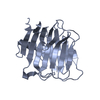 1c4rS S: Starting model for refinement C: citing same article ( |
|---|---|
| Similar structure data |


-Links
 PDBj
PDBj


- Assembly
| Deposited unit | 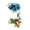
| ||||||||
|---|---|---|---|---|---|---|---|---|---|
| 1 |
| ||||||||
| Unit cell |
|
-Components
| #1: Protein | Mass: 44838.309 Da / Num. of mol.: 1 / Fragment: UNP residues 923-1323 Source method: isolated from a genetically manipulated source Source: (gene. exp.)  Cricetulus griseus (Chinese hamster) / References: UniProt: Q28146 Cricetulus griseus (Chinese hamster) / References: UniProt: Q28146 |
|---|---|
| #2: Chemical | ChemComp-CA /   Mass: 40.078 Da / Num. of mol.: 1 / Source method: obtained synthetically / Formula: Ca Mass: 40.078 Da / Num. of mol.: 1 / Source method: obtained synthetically / Formula: Ca |
| #3: Sugar | ChemComp-NAG /   Type: D-saccharide, beta linking / Mass: 221.208 Da / Num. of mol.: 1 Type: D-saccharide, beta linking / Mass: 221.208 Da / Num. of mol.: 1Source method: isolated from a genetically manipulated source Formula: C8H15NO6 |
| #4: Water | ChemComp-HOH /  Mass: 18.015 Da / Num. of mol.: 148 / Source method: isolated from a natural source / Formula: H2O Mass: 18.015 Da / Num. of mol.: 148 / Source method: isolated from a natural source / Formula: H2O |
| Has protein modification | Y |
| Sequence details | 1. RESIDUE 1068 (UNP RESIDUE NUMBER 1128) WAS A PHE IN THE CDNA USED FOR THIS STUDY; 2. SEQUENCE OF ...1. RESIDUE 1068 (UNP RESIDUE NUMBER 1128) WAS A PHE IN THE CDNA USED FOR THIS STUDY; 2. SEQUENCE OF THE PROTEIN WAS BASED ON ISOFORM 11 OF DATABASE Q28146 (NRX1A_BOVIN). |
-Experimental details
-Experiment
| Experiment | Method: X-RAY DIFFRACTION / Number of used crystals: 1 |
|---|
- Sample preparation
| Crystal | Density Matthews: 2.52 Å3/Da / Density % sol: 51.22 % |
|---|---|
| Crystal grow | Temperature: 293 K / Method: vapor diffusion, hanging drop / pH: 7 Details: 18% PEG3000, 0.1M sodium acetate and 0.3M sodium malonate pH 7.0, VAPOR DIFFUSION, HANGING DROP, temperature 293KK |
-Data collection
| Diffraction | Mean temperature: 100 K |
|---|---|
| Diffraction source | Source: SYNCHROTRON / Site: SPring-8 / Beamline: BL41XU / Wavelength: 1 Å |
| Detector | Type: ADSC QUANTUM 315r / Detector: CCD / Date: Oct 16, 2006 |
| Radiation | Monochromator: rotated-inclined double-crystal monochromator, Si(111) Protocol: SINGLE WAVELENGTH / Monochromatic (M) / Laue (L): M / Scattering type: x-ray |
| Radiation wavelength | Wavelength: 1 Å / Relative weight: 1 |
| Reflection | Resolution: 2.3→39.07 Å / Num. all: 20732 / Num. obs: 20732 / % possible obs: 99.9 % / Observed criterion σ(F): 0 / Observed criterion σ(I): -3 / Redundancy: 7.1 % / Biso Wilson estimate: 28.8 Å2 / Rmerge(I) obs: 0.083 / Net I/σ(I): 20.9 |
| Reflection shell | Resolution: 2.3→2.42 Å / Redundancy: 7.2 % / Rmerge(I) obs: 0.271 / Mean I/σ(I) obs: 6.2 / Num. unique all: 2962 / % possible all: 100 |
- Processing
| Software |
| |||||||||||||||||||||||||||||||||||||||||||||||||||||||||||||||||
|---|---|---|---|---|---|---|---|---|---|---|---|---|---|---|---|---|---|---|---|---|---|---|---|---|---|---|---|---|---|---|---|---|---|---|---|---|---|---|---|---|---|---|---|---|---|---|---|---|---|---|---|---|---|---|---|---|---|---|---|---|---|---|---|---|---|---|
| Refinement | Method to determine structure: MOLECULAR REPLACEMENT Starting model: PDB ENTRY 1C4R Resolution: 2.3→39.07 Å / Cor.coef. Fo:Fc: 0.94 / Cor.coef. Fo:Fc free: 0.903 / SU B: 6.079 / SU ML: 0.152 / Cross valid method: THROUGHOUT / ESU R Free: 0.233 / Stereochemistry target values: MAXIMUM LIKELIHOOD
| |||||||||||||||||||||||||||||||||||||||||||||||||||||||||||||||||
| Solvent computation | Ion probe radii: 0.8 Å / Shrinkage radii: 0.8 Å / VDW probe radii: 1.2 Å / Solvent model: MASK | |||||||||||||||||||||||||||||||||||||||||||||||||||||||||||||||||
| Displacement parameters | Biso mean: 24.548 Å2
| |||||||||||||||||||||||||||||||||||||||||||||||||||||||||||||||||
| Refinement step | Cycle: LAST / Resolution: 2.3→39.07 Å
| |||||||||||||||||||||||||||||||||||||||||||||||||||||||||||||||||
| Refine LS restraints |
| |||||||||||||||||||||||||||||||||||||||||||||||||||||||||||||||||
| LS refinement shell | Resolution: 2.3→2.36 Å / Total num. of bins used: 20
|
