 Movie
Movie Controller


+ Open data

- Basic information
| Entry | Database: PDB / ID: 3amh | ||||||
|---|---|---|---|---|---|---|---|
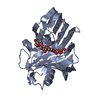
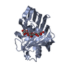
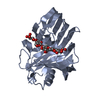
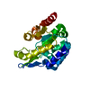
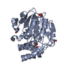
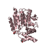
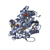
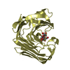
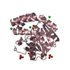
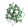
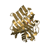
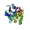
| Title | crystal structure of cellulase 12A from Thermotoga maritima | ||||||
 Components Components | Endo-1,4-beta-glucanase | ||||||
 Keywords Keywords | HYDROLASE / beta jellyroll / glucanase / cellulose | ||||||
| Function / homology |  Function and homology information Function and homology information | ||||||
| Biological species |   Thermotoga maritima (bacteria) Thermotoga maritima (bacteria) | ||||||
| Method |  X-RAY DIFFRACTION / SYNCHROTRON / MIR / Resolution: 2.09 Å X-RAY DIFFRACTION / SYNCHROTRON / MIR / Resolution: 2.09 Å | ||||||
 Authors Authors | Cheng, Y.-S. / Ko, T.-P. / Liu, J.-R. / Guo, R.-T. | ||||||
 Citation Citation | Journal: Proteins / Year: 2011 Title: Crystal structure and substrate-binding mode of cellulase 12A from Thermotoga maritima Authors: Cheng, Y.-S. / Ko, T.-P. / Wu, T.-H. / Ma, Y. / Huang, C.-H. / Lai, H.-L. / Wang, A.H.-J. / Liu, J.-R. / Guo, R.-T. | ||||||
| History |
|
- Structure visualization
| Structure viewer | Molecule: MolmilJmol/JSmol |
|---|
- Downloads & links
-Download
| PDBx/mmCIF format | 3amh.cif.gz | 121.3 KB | Display | PDBx/mmCIF format |
|---|---|---|---|---|
| PDB format | pdb3amh.ent.gz | 95 KB | Display | PDB format |
| PDBx/mmJSON format | 3amh.json.gz | Tree view | PDBx/mmJSON format | |
| Others |  Other downloads Other downloads |
-Validation report
| Summary document | 3amh_validation.pdf.gz | 435.1 KB | Display | wwPDB validaton report |
|---|---|---|---|---|
| Full document | 3amh_full_validation.pdf.gz | 441.2 KB | Display | |
| Data in XML | 3amh_validation.xml.gz | 23.5 KB | Display | |
| Data in CIF | 3amh_validation.cif.gz | 33.9 KB | Display | |
| Arichive directory | https://data.pdbj.org/pub/pdb/validation_reports/am/3amhftp://data.pdbj.org/pub/pdb/validation_reports/am/3amh | HTTPS FTP |
-Related structure data


-Links
 PDBj
PDBj
- Assembly
| Deposited unit | 
| ||||||||
|---|---|---|---|---|---|---|---|---|---|
| 1 | 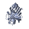
| ||||||||
| 2 | 
| ||||||||
| Unit cell |
|
-Components
| #1: Protein | Mass: 30779.611 Da / Num. of mol.: 2 Source method: isolated from a genetically manipulated source Source: (gene. exp.) Thermotoga maritima (bacteria) / Gene: celA / Plasmid: pET16b / Production host: #2: Water | ChemComp-HOH / |  Mass: 18.015 Da / Num. of mol.: 296 / Source method: isolated from a natural source / Formula: H2O Mass: 18.015 Da / Num. of mol.: 296 / Source method: isolated from a natural source / Formula: H2O |
|---|
-Experimental details
-Experiment
| Experiment | Method: X-RAY DIFFRACTION / Number of used crystals: 1 |
|---|
- Sample preparation
| Crystal | Density Matthews: 2.27 Å3/Da / Density % sol: 45.74 % |
|---|---|
| Crystal grow | Temperature: 298 K / Method: vapor diffusion, sitting drop / pH: 5.5 Details: 0.1M Bis-Tris, 10% glycerol, 15% PEG3350, pH 5.5, VAPOR DIFFUSION, SITTING DROP, temperature 298K |
-Data collection
| Diffraction | Mean temperature: 100 K |
|---|---|
| Diffraction source | Source: SYNCHROTRON / Site: NSRRC  / Beamline: BL13B1 / Wavelength: 0.9732 Å / Beamline: BL13B1 / Wavelength: 0.9732 Å |
| Detector | Type: ADSC QUANTUM 315r / Detector: CCD / Date: Sep 24, 2009 |
| Radiation | Monochromator: Si 111 CHANNEL / Protocol: SINGLE WAVELENGTH / Monochromatic (M) / Laue (L): M / Scattering type: x-ray |
| Radiation wavelength | Wavelength: 0.9732 Å / Relative weight: 1 |
| Reflection | Resolution: 2.09→25 Å / Num. all: 34200 / Num. obs: 33848 / % possible obs: 98.7 % / Observed criterion σ(F): 0 / Observed criterion σ(I): 3 / Redundancy: 5.4 % / Rmerge(I) obs: 0.103 / Net I/σ(I): 20.2 |
| Reflection shell | Resolution: 2.09→2.16 Å / Redundancy: 5 % / Rmerge(I) obs: 0.293 / Mean I/σ(I) obs: 6.8 / % possible all: 99.6 |
- Processing
| Software |
| ||||||||||||||||||||
|---|---|---|---|---|---|---|---|---|---|---|---|---|---|---|---|---|---|---|---|---|---|
| Refinement | Method to determine structure: MIR / Resolution: 2.09→25 Å / σ(F): 0 / Stereochemistry target values: Engh & Huber
| ||||||||||||||||||||
| Refine analyze |
| ||||||||||||||||||||
| Refinement step | Cycle: LAST / Resolution: 2.09→25 Å
| ||||||||||||||||||||
| Refine LS restraints |
|
