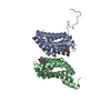
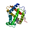
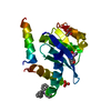
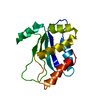
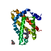
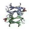
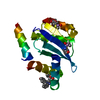
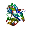
- PDB-3ag0: Crystal structure of carbonmonoxy human cytoglobin -
+
Open data
ID or keywords:
Loading...
-
Basic information
Entry
Database: PDB / ID: 3ag0
Title
Crystal structure of carbonmonoxy human cytoglobin
Components
Cytoglobin
Keywords
OXYGEN TRANSPORT / Globins / Heme / Carbon Monoxide / Complex / Ligands / hexacoordination / bis-His / Disulfide bond / Iron / Metal-binding / Transport
Function / homology
Function and homology information
fatty acid peroxidase activity / nitric oxide dioxygenase activity, heme protein as donor / negative regulation of hepatic stellate cell activation / Oxidoreductases; Acting on paired donors, with incorporation or reduction of molecular oxygen; With NADH or NADPH as one donor, and incorporation of two atoms of oxygen into the other donor / Intracellular oxygen transport / nitric oxide catabolic process / carbon monoxide binding / negative regulation of collagen biosynthetic process / negative regulation of fibroblast migration / catalase activity ...fatty acid peroxidase activity / nitric oxide dioxygenase activity, heme protein as donor / negative regulation of hepatic stellate cell activation / Oxidoreductases; Acting on paired donors, with incorporation or reduction of molecular oxygen; With NADH or NADPH as one donor, and incorporation of two atoms of oxygen into the other donor / Intracellular oxygen transport / nitric oxide catabolic process / carbon monoxide binding / negative regulation of collagen biosynthetic process / negative regulation of fibroblast migration / catalase activity / superoxide dismutase / superoxide dismutase activity / Oxidoreductases; Acting on other nitrogenous compounds as donors / nitrite reductase activity / fatty acid oxidation / oxygen transport / Oxidoreductases; Acting on a peroxide as acceptor; Peroxidases / nitric oxide metabolic process / eNOS activation / removal of superoxide radicals / oxygen carrier activity / peroxidase activity / oxygen binding / response to oxidative stress / response to hypoxia / oxidoreductase activity / neuron projection / nuclear speck / iron ion binding / neuronal cell body / heme binding / nucleus / cytoplasm / cytosol Similarity search - Function
In the structure databanks used in Yorodumi, some data are registered as the other names, "COVID-19 virus" and "2019-nCoV". Here are the details of the virus and the list of structure data.
Jan 31, 2019. EMDB accession codes are about to change! (news from PDBe EMDB page)
EMDB accession codes are about to change! (news from PDBe EMDB page)
The allocation of 4 digits for EMDB accession codes will soon come to an end. Whilst these codes will remain in use, new EMDB accession codes will include an additional digit and will expand incrementally as the available range of codes is exhausted. The current 4-digit format prefixed with “EMD-” (i.e. EMD-XXXX) will advance to a 5-digit format (i.e. EMD-XXXXX), and so on. It is currently estimated that the 4-digit codes will be depleted around Spring 2019, at which point the 5-digit format will come into force.
The EM Navigator/Yorodumi systems omit the EMD- prefix.
Related info.:Q: What is EMD? / ID/Accession-code notation in Yorodumi/EM Navigator
Yorodumi is a browser for structure data from EMDB, PDB, SASBDB, etc.
This page is also the successor to EM Navigator detail page, and also detail information page/front-end page for Omokage search.
The word "yorodu" (or yorozu) is an old Japanese word meaning "ten thousand". "mi" (miru) is to see.
Related info.:EMDB / PDB / SASBDB / Comparison of 3 databanks / Yorodumi Search / Aug 31, 2016. New EM Navigator & Yorodumi / Yorodumi Papers / Jmol/JSmol / Function and homology information / Changes in new EM Navigator and Yorodumi
 Movie
Movie Controller


 Open data
 Basic information
 Components
 Keywords
 Function and homology information
 Homo sapiens (human)
 X-RAY DIFFRACTION /
  Authors
 Citation
 Structure visualization
 Downloads & links
 Other downloads
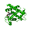

 PDBj
PDBj


 Assembly


 Mass: 616.487 Da / Num. of mol.: 1 / Source method: obtained synthetically / Formula: C34H32FeN4O4
 Mass: 28.010 Da / Num. of mol.: 1 / Source method: obtained synthetically / Formula: CO
 Mass: 18.015 Da / Num. of mol.: 43 / Source method: isolated from a natural source / Formula: H2O
 Sample preparation
 / Beamline: BL41XU / Wavelength: 1 Å
 Processing