[English] 日本語
 Yorodumi
- PDB-3afp: Crystal structure of the single-stranded DNA binding protein from... -
+ Open data
- Basic information
| Entry | Database: PDB / ID: 3afp | ||||||
|---|---|---|---|---|---|---|---|
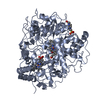
| Title | Crystal structure of the single-stranded DNA binding protein from Mycobacterium leprae (Form I) | ||||||
 Components Components | Single-stranded DNA-binding protein | ||||||
 Keywords Keywords | DNA BINDING PROTEIN / OB-fold / quaternary structure and stability / changes on oligomerisation / water-bridges / DNA damage / DNA repair / DNA replication / DNA-binding | ||||||
| Function / homology |  Function and homology information Function and homology information | ||||||
| Biological species |  Mycobacterium leprae (bacteria) Mycobacterium leprae (bacteria) | ||||||
| Method |  X-RAY DIFFRACTION / X-RAY DIFFRACTION /  MOLECULAR REPLACEMENT / Resolution: 2.05 Å MOLECULAR REPLACEMENT / Resolution: 2.05 Å | ||||||
 Authors Authors | Kaushal, P.S. / Singh, P. / Sharma, A. / Muniyappa, K. / Vijayan, M. | ||||||
 Citation Citation |  Journal: Acta Crystallogr.,Sect.D / Year: 2010 Journal: Acta Crystallogr.,Sect.D / Year: 2010Title: X-ray and molecular-dynamics studies on Mycobacterium leprae single-stranded DNA-binding protein and comparison with other eubacterial SSB structures Authors: Kaushal, P.S. / Singh, P. / Sharma, A. / Muniyappa, K. / Vijayan, M. #1:  Journal: Acta Crystallogr.,Sect.D / Year: 2005 Journal: Acta Crystallogr.,Sect.D / Year: 2005Title: Structure of Mycobacterium smegmatis single-stranded DNA-binding protein and a comparative study involving homologus SSBs: biological implications of structural plasticity and variability in quaternary association Authors: Saikrishnan, K. / Manjunath, G.P. / Singh, P. / Jeyakanthan, J. / Dauter, Z. / Sekar, K. / Muniyappa, K. / Vijayan, M. #2:  Journal: J.Mol.Biol. / Year: 2003 Journal: J.Mol.Biol. / Year: 2003Title: Structure of Mycobacterium tuberculosis single-stranded DNA-binding protein. Variability in quaternary structure and its implications Authors: Saikrishnan, K. / Jeyakanthan, J. / Venkatesh, J. / Acharya, N. / Sekar, K. / Varshney, U. / Vijayan, M. #3:  Journal: Nat.Struct.Biol. / Year: 1997 Journal: Nat.Struct.Biol. / Year: 1997Title: Crystal structure of human mitochondrial single-stranded DNA binding protein at 2.4 A resolution Authors: Yang, C. / Curth, U. / Urbanke, C. / Kang, C. #4:  Journal: Proc.Natl.Acad.Sci.USA / Year: 1997 Journal: Proc.Natl.Acad.Sci.USA / Year: 1997Title: Crystal structure of the homo-tetrameric DNA binding domain of Escherichia coli single-stranded DNA-binding protein determined by multiwavelength x-ray diffraction on the selenomethionyl ...Title: Crystal structure of the homo-tetrameric DNA binding domain of Escherichia coli single-stranded DNA-binding protein determined by multiwavelength x-ray diffraction on the selenomethionyl protein at 2.9-A resolution Authors: Raghunathan, S. / Ricard, C.S. / Lohman, T.M. / Waksman, G. | ||||||
| History |
|
- Structure visualization
| Structure viewer | Molecule:  Molmil Molmil Jmol/JSmol Jmol/JSmol |
|---|
- Downloads & links
- Download
| PDBx/mmCIF format |  3afp.cif.gz 3afp.cif.gz | 105.2 KB | Display |  PDBx/mmCIF format PDBx/mmCIF format |
|---|---|---|---|---|
| PDB format |  pdb3afp.ent.gz pdb3afp.ent.gz | 80 KB | Display |  PDB format PDB format |
| PDBx/mmJSON format |  3afp.json.gz 3afp.json.gz | Tree view |  PDBx/mmJSON format PDBx/mmJSON format | |
| Others |  Other downloads Other downloads |
-Validation report
| Summary document |  3afp_validation.pdf.gz 3afp_validation.pdf.gz | 450.2 KB | Display |  wwPDB validaton report wwPDB validaton report |
|---|---|---|---|---|
| Full document |  3afp_full_validation.pdf.gz 3afp_full_validation.pdf.gz | 454.8 KB | Display | |
| Data in XML |  3afp_validation.xml.gz 3afp_validation.xml.gz | 13.6 KB | Display | |
| Data in CIF |  3afp_validation.cif.gz 3afp_validation.cif.gz | 19.4 KB | Display | |
| Arichive directory |  https://data.pdbj.org/pub/pdb/validation_reports/af/3afp https://data.pdbj.org/pub/pdb/validation_reports/af/3afp ftp://data.pdbj.org/pub/pdb/validation_reports/af/3afp ftp://data.pdbj.org/pub/pdb/validation_reports/af/3afp | HTTPS FTP |
-Related structure data
| Related structure data |  3afqC  1x3eS C: citing same article ( S: Starting model for refinement |
|---|---|
| Similar structure data |
- Links
Links
- Assembly
| Deposited unit | 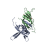
| ||||||||||||||||||
|---|---|---|---|---|---|---|---|---|---|---|---|---|---|---|---|---|---|---|---|
| 1 | 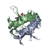
| ||||||||||||||||||
| Unit cell |
| ||||||||||||||||||
| Components on special symmetry positions |
|
- Components
| #1: Protein | Mass: 17719.500 Da / Num. of mol.: 2 Source method: isolated from a genetically manipulated source Source: (gene. exp.)  Mycobacterium leprae (bacteria) / Strain: TN / Gene: ssb / Plasmid: pET21a, pMLSSB / Production host: Mycobacterium leprae (bacteria) / Strain: TN / Gene: ssb / Plasmid: pET21a, pMLSSB / Production host:  #2: Chemical | ChemComp-GOL / | #3: Chemical | ChemComp-CD / | #4: Water | ChemComp-HOH / | |
|---|
-Experimental details
-Experiment
| Experiment | Method:  X-RAY DIFFRACTION / Number of used crystals: 1 X-RAY DIFFRACTION / Number of used crystals: 1 |
|---|
- Sample preparation
| Crystal | Density Matthews: 2.04 Å3/Da / Density % sol: 39.61 % |
|---|---|
| Crystal grow | Temperature: 298 K / Method: vapor diffusion, hanging drop / pH: 7.5 Details: 1M Sodium acetate, 500mM NaCl, 30mM CdSO4, 20mM Tris-HCl, pH 7.5, VAPOR DIFFUSION, HANGING DROP, temperature 298K |
-Data collection
| Diffraction | Mean temperature: 100 K |
|---|---|
| Diffraction source | Source:  ROTATING ANODE / Type: RIGAKU RU200 / Wavelength: 1.5418 Å ROTATING ANODE / Type: RIGAKU RU200 / Wavelength: 1.5418 Å |
| Detector | Type: MAR scanner 345 mm plate / Detector: IMAGE PLATE / Date: Jun 27, 2006 / Details: mirrors |
| Radiation | Monochromator: osmic mirror / Protocol: SINGLE WAVELENGTH / Monochromatic (M) / Laue (L): M / Scattering type: x-ray |
| Radiation wavelength | Wavelength: 1.5418 Å / Relative weight: 1 |
| Reflection | Resolution: 2.05→30 Å / Num. obs: 18540 / % possible obs: 99.8 % / Observed criterion σ(I): 0 / Redundancy: 3.9 % / Biso Wilson estimate: 48.3 Å2 / Rsym value: 0.06 / Net I/σ(I): 21.6 |
| Reflection shell | Resolution: 2.05→2.12 Å / Mean I/σ(I) obs: 3.1 / Num. unique all: 1824 / Rsym value: 0.461 / % possible all: 99.9 |
- Processing
| Software |
| |||||||||||||||||||||||||||||||||||||||||||||||||||||||||||||||||||||||||||||||||||||||||||||||||||||||||||||||||||||||||||||||||||||||||||||||||||||||||||||||||||||||||||||||||||||||||||||||||||||||||||||||||||||||||||||||||
|---|---|---|---|---|---|---|---|---|---|---|---|---|---|---|---|---|---|---|---|---|---|---|---|---|---|---|---|---|---|---|---|---|---|---|---|---|---|---|---|---|---|---|---|---|---|---|---|---|---|---|---|---|---|---|---|---|---|---|---|---|---|---|---|---|---|---|---|---|---|---|---|---|---|---|---|---|---|---|---|---|---|---|---|---|---|---|---|---|---|---|---|---|---|---|---|---|---|---|---|---|---|---|---|---|---|---|---|---|---|---|---|---|---|---|---|---|---|---|---|---|---|---|---|---|---|---|---|---|---|---|---|---|---|---|---|---|---|---|---|---|---|---|---|---|---|---|---|---|---|---|---|---|---|---|---|---|---|---|---|---|---|---|---|---|---|---|---|---|---|---|---|---|---|---|---|---|---|---|---|---|---|---|---|---|---|---|---|---|---|---|---|---|---|---|---|---|---|---|---|---|---|---|---|---|---|---|---|---|---|---|---|---|---|---|---|---|---|---|---|---|---|---|---|---|---|---|
| Refinement | Method to determine structure:  MOLECULAR REPLACEMENT MOLECULAR REPLACEMENTStarting model: PDB ENTRY 1X3E Resolution: 2.05→28.14 Å / Cor.coef. Fo:Fc: 0.954 / Cor.coef. Fo:Fc free: 0.937 / SU B: 8.454 / SU ML: 0.112 / Cross valid method: THROUGHOUT / σ(F): 0 / ESU R: 0.187 / ESU R Free: 0.159 / Stereochemistry target values: MAXIMUM LIKELIHOOD / Details: Bulak solvent model user
| |||||||||||||||||||||||||||||||||||||||||||||||||||||||||||||||||||||||||||||||||||||||||||||||||||||||||||||||||||||||||||||||||||||||||||||||||||||||||||||||||||||||||||||||||||||||||||||||||||||||||||||||||||||||||||||||||
| Solvent computation | Ion probe radii: 0.8 Å / Shrinkage radii: 0.8 Å / VDW probe radii: 1.4 Å / Solvent model: MASK | |||||||||||||||||||||||||||||||||||||||||||||||||||||||||||||||||||||||||||||||||||||||||||||||||||||||||||||||||||||||||||||||||||||||||||||||||||||||||||||||||||||||||||||||||||||||||||||||||||||||||||||||||||||||||||||||||
| Displacement parameters | Biso mean: 49.809 Å2
| |||||||||||||||||||||||||||||||||||||||||||||||||||||||||||||||||||||||||||||||||||||||||||||||||||||||||||||||||||||||||||||||||||||||||||||||||||||||||||||||||||||||||||||||||||||||||||||||||||||||||||||||||||||||||||||||||
| Refinement step | Cycle: LAST / Resolution: 2.05→28.14 Å
| |||||||||||||||||||||||||||||||||||||||||||||||||||||||||||||||||||||||||||||||||||||||||||||||||||||||||||||||||||||||||||||||||||||||||||||||||||||||||||||||||||||||||||||||||||||||||||||||||||||||||||||||||||||||||||||||||
| Refine LS restraints |
| |||||||||||||||||||||||||||||||||||||||||||||||||||||||||||||||||||||||||||||||||||||||||||||||||||||||||||||||||||||||||||||||||||||||||||||||||||||||||||||||||||||||||||||||||||||||||||||||||||||||||||||||||||||||||||||||||
| LS refinement shell | Resolution: 2.05→2.103 Å / Total num. of bins used: 20
| |||||||||||||||||||||||||||||||||||||||||||||||||||||||||||||||||||||||||||||||||||||||||||||||||||||||||||||||||||||||||||||||||||||||||||||||||||||||||||||||||||||||||||||||||||||||||||||||||||||||||||||||||||||||||||||||||
| Refinement TLS params. | Method: refined / Refine-ID: X-RAY DIFFRACTION
| |||||||||||||||||||||||||||||||||||||||||||||||||||||||||||||||||||||||||||||||||||||||||||||||||||||||||||||||||||||||||||||||||||||||||||||||||||||||||||||||||||||||||||||||||||||||||||||||||||||||||||||||||||||||||||||||||
| Refinement TLS group |
|
 Movie
Movie Controller






 PDBj
PDBj


