 Movie
Movie Controller

[English] 日本語
 Yorodumi
- PDB-3ach: Crystal Structure of Carbohydrate-Binding Module Family 28 from C... -
+ Open data

- Basic information
| Entry | Database: PDB / ID: 3ach | |||||||||
|---|---|---|---|---|---|---|---|---|---|---|
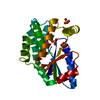
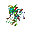
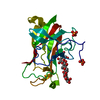
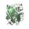
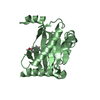
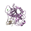
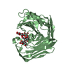
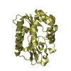
| Title | Crystal Structure of Carbohydrate-Binding Module Family 28 from Clostridium josui Cel5A in complex with cellotetraose | |||||||||
 Components Components | Beta-1,4-endoglucanase | |||||||||
 Keywords Keywords | HYDROLASE / beta-jellyroll / cellulose-binding domain | |||||||||
| Function / homology |  Function and homology information Function and homology informationcellulase / cellulase activity / cellulose catabolic process / metal ion binding Similarity search - Function | |||||||||
| Biological species |  Clostridium josui (bacteria) Clostridium josui (bacteria) | |||||||||
| Method |  X-RAY DIFFRACTION / SYNCHROTRON / MOLECULAR REPLACEMENT / Resolution: 1.4 Å X-RAY DIFFRACTION / SYNCHROTRON / MOLECULAR REPLACEMENT / Resolution: 1.4 Å | |||||||||
 Authors Authors | Tsukimoto, K. / Takada, R. / Araki, Y. / Suzuki, K. / Karita, S. / Wakagi, T. / Shoun, H. / Watanabe, T. / Fushinobu, S. | |||||||||
 Citation Citation | Journal: Febs Lett. / Year: 2010 Title: Recognition of cellooligosaccharides by a family 28 carbohydrate-binding module. Authors: Tsukimoto, K. / Takada, R. / Araki, Y. / Suzuki, K. / Karita, S. / Wakagi, T. / Shoun, H. / Watanabe, T. / Fushinobu, S. | |||||||||
| History |
|
- Structure visualization
| Structure viewer | Molecule: MolmilJmol/JSmol |
|---|
- Downloads & links
-Download
| PDBx/mmCIF format | 3ach.cif.gz | 63.5 KB | Display | PDBx/mmCIF format |
|---|---|---|---|---|
| PDB format | pdb3ach.ent.gz | 43.7 KB | Display | PDB format |
| PDBx/mmJSON format | 3ach.json.gz | Tree view | PDBx/mmJSON format | |
| Others |  Other downloads Other downloads |
-Validation report
| Arichive directory | https://data.pdbj.org/pub/pdb/validation_reports/ac/3achftp://data.pdbj.org/pub/pdb/validation_reports/ac/3ach | HTTPS FTP |
|---|
-Related structure data
| Related structure data | 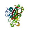 3acfC  3acgC  3aciC 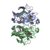 1uwwS C: citing same article ( S: Starting model for refinement |
|---|---|
| Similar structure data |


-Links
 PDBj
PDBj- Assembly
| Deposited unit | 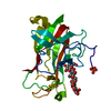
| ||||||||
|---|---|---|---|---|---|---|---|---|---|
| 1 |
| ||||||||
| Unit cell |
|
-Components
| #1: Protein | Mass: 22390.189 Da / Num. of mol.: 1 / Fragment: UNP residues 560-752 Source method: isolated from a genetically manipulated source Source: (gene. exp.) Clostridium josui (bacteria) / Gene: celA / Plasmid: pQE30 / Production host: |
|---|---|
| #2: Polysaccharide | beta-D-glucopyranose-(1-4)-beta-D-glucopyranose-(1-4)-beta-D-glucopyranose-(1-4)-beta-D-glucopyranose / beta-cellotetraose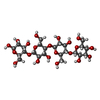  Source method: isolated from a genetically manipulated source Details: oligosaccharide / References: beta-cellotetraose |
| #3: Chemical | ChemComp-CA /   Mass: 40.078 Da / Num. of mol.: 1 / Source method: obtained synthetically / Formula: Ca Mass: 40.078 Da / Num. of mol.: 1 / Source method: obtained synthetically / Formula: Ca |
| #4: Chemical | ChemComp-PO4 /   Mass: 94.971 Da / Num. of mol.: 1 / Source method: obtained synthetically / Formula: PO4 Mass: 94.971 Da / Num. of mol.: 1 / Source method: obtained synthetically / Formula: PO4 |
| #5: Water | ChemComp-HOH /  Mass: 18.015 Da / Num. of mol.: 356 / Source method: isolated from a natural source / Formula: H2O Mass: 18.015 Da / Num. of mol.: 356 / Source method: isolated from a natural source / Formula: H2O |
-Experimental details
-Experiment
| Experiment | Method: X-RAY DIFFRACTION / Number of used crystals: 1 |
|---|
- Sample preparation
| Crystal | Density Matthews: 2.14 Å3/Da / Density % sol: 42.39 % |
|---|---|
| Crystal grow | Temperature: 277 K / Method: vapor diffusion, sitting drop Details: PEG 8000, KH2PO4, vapor diffusion, sitting drop, temperature 277K |
-Data collection
| Diffraction | Mean temperature: 100 K |
|---|---|
| Diffraction source | Source: SYNCHROTRON / Site: Photon Factory  / Beamline: BL-5A / Wavelength: 1 Å / Beamline: BL-5A / Wavelength: 1 Å |
| Detector | Type: ADSC QUANTUM 315 / Detector: CCD / Date: Dec 1, 2009 |
| Radiation | Protocol: SINGLE WAVELENGTH / Monochromatic (M) / Laue (L): M / Scattering type: x-ray |
| Radiation wavelength | Wavelength: 1 Å / Relative weight: 1 |
| Reflection | Resolution: 1.4→50 Å / Num. obs: 35569 / % possible obs: 92.3 % / Observed criterion σ(I): 0 / Redundancy: 6.3 % / Rsym value: 0.047 / Net I/σ(I): 38.7 |
| Reflection shell | Resolution: 1.4→1.42 Å / Redundancy: 6.4 % / Mean I/σ(I) obs: 5.2 / Rsym value: 0.247 / % possible all: 95.5 |
- Processing
| Software |
| |||||||||||||||||||||||||||||||||||||||||||||||||||||||||||||||||
|---|---|---|---|---|---|---|---|---|---|---|---|---|---|---|---|---|---|---|---|---|---|---|---|---|---|---|---|---|---|---|---|---|---|---|---|---|---|---|---|---|---|---|---|---|---|---|---|---|---|---|---|---|---|---|---|---|---|---|---|---|---|---|---|---|---|---|
| Refinement | Method to determine structure: MOLECULAR REPLACEMENT Starting model: 1UWW Resolution: 1.4→27.34 Å / Cor.coef. Fo:Fc: 0.967 / Cor.coef. Fo:Fc free: 0.956 / Occupancy max: 1 / Occupancy min: 1 / SU B: 0.894 / SU ML: 0.037 / Cross valid method: THROUGHOUT / σ(F): 0 / ESU R: 0.064 / ESU R Free: 0.069 / Stereochemistry target values: MAXIMUM LIKELIHOOD Details: HYDROGENS HAVE BEEN ADDED IN THE RIDING POSITIONS U VALUES: REFINED INDIVIDUALLY
| |||||||||||||||||||||||||||||||||||||||||||||||||||||||||||||||||
| Solvent computation | Ion probe radii: 0.8 Å / Shrinkage radii: 0.8 Å / VDW probe radii: 1.4 Å / Solvent model: BABINET MODEL WITH MASK | |||||||||||||||||||||||||||||||||||||||||||||||||||||||||||||||||
| Displacement parameters | Biso max: 55.44 Å2 / Biso mean: 19.325 Å2 / Biso min: 9.57 Å2
| |||||||||||||||||||||||||||||||||||||||||||||||||||||||||||||||||
| Refine analyze |
| |||||||||||||||||||||||||||||||||||||||||||||||||||||||||||||||||
| Refinement step | Cycle: LAST / Resolution: 1.4→27.34 Å
| |||||||||||||||||||||||||||||||||||||||||||||||||||||||||||||||||
| Refine LS restraints |
| |||||||||||||||||||||||||||||||||||||||||||||||||||||||||||||||||
| LS refinement shell | Resolution: 1.401→1.437 Å / Total num. of bins used: 20
|
