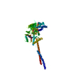
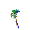
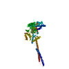
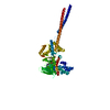
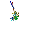
Entry Database : PDB / ID : 3abtTitle Crystal Structure of LSD1 in complex with trans-2-pentafluorophenylcyclopropylamine Lysine-specific histone demethylase 1 Keywords / / / / / / / / / / / / / / / / / Function / homology Function Domain/homology Component
/ / / / / / / / / / / / / / / / / / / / / / / / / / / / / / / / / / / / / / / / / / / / / / / / / / / / / / / / / / / / / / / / / / / / / / / / / / / / / / / / / / / / / / / / / / / / / / / / / / / / Biological species Homo sapiens (human)Method / / / Resolution : 3.2 Å Authors Mimasu, S. / Umezawa, N. / Sato, S. / Higuchi, T. / Umehara, T. / Yokoyama, S. / RIKEN Structural Genomics/Proteomics Initiative (RSGI) Journal : Biochemistry / Year : 2010Title : Structurally Designed trans-2-Phenylcyclopropylamine Derivatives Potently Inhibit Histone Demethylase LSD1/KDM1Authors : Mimasu, S. / Umezawa, N. / Sato, S. / Higuchi, T. / Umehara, T. / Yokoyama, S. History Deposition Dec 21, 2009 Deposition site / Processing site Revision 1.0 Jul 7, 2010 Provider / Type Revision 1.1 Jul 13, 2011 Group Revision 1.2 Nov 1, 2023 Group Data collection / Database references ... Data collection / Database references / Derived calculations / Refinement description Category chem_comp_atom / chem_comp_bond ... chem_comp_atom / chem_comp_bond / database_2 / pdbx_initial_refinement_model / struct_site Item _database_2.pdbx_DOI / _database_2.pdbx_database_accession ... _database_2.pdbx_DOI / _database_2.pdbx_database_accession / _struct_site.pdbx_auth_asym_id / _struct_site.pdbx_auth_comp_id / _struct_site.pdbx_auth_seq_id
Show all Show less
 Movie
Movie Controller

 Yorodumi
 Open data
 Basic information
 Components
 Keywords
 Function and homology information
 Homo sapiens (human)
 X-RAY DIFFRACTION /
  Authors
 Citation
 Structure visualization
 Downloads & links
 Other downloads


 PDBj
PDBj


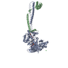
 Assembly
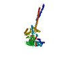

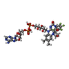
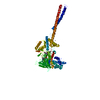
 Mass: 1009.677 Da / Num. of mol.: 1 / Source method: obtained synthetically / Formula: C36H38F5N9O16P2
 Sample preparation
 Processing