 Movie
Movie Controller

[English] 日本語
 Yorodumi
- PDB-3a8h: Crystal structure of Nitrile Hydratase mutant S113A complexed wit... -
+ Open data

- Basic information
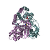
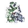
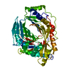
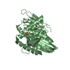
| Entry | Database: PDB / ID: 3a8h | ||||||
|---|---|---|---|---|---|---|---|
| Title | Crystal structure of Nitrile Hydratase mutant S113A complexed with Trimethylacetamide | ||||||
 Components Components |
| ||||||
 Keywords Keywords | LYASE / nitrile hydratase / Fe / Iron / Metal-binding / Oxidation | ||||||
| Function / homology |  Function and homology information Function and homology informationnitrile hydratase / nitrile hydratase activity / transition metal ion binding Similarity search - Function | ||||||
| Biological species |  Rhodococcus erythropolis (bacteria) Rhodococcus erythropolis (bacteria) | ||||||
| Method |  X-RAY DIFFRACTION / SYNCHROTRON / MOLECULAR REPLACEMENT / Resolution: 1.66 Å X-RAY DIFFRACTION / SYNCHROTRON / MOLECULAR REPLACEMENT / Resolution: 1.66 Å | ||||||
 Authors Authors | Yamanaka, Y. / Hashimoto, K. / Ohtaki, A. / Noguchi, K. / Yohda, M. / Odaka, M. | ||||||
 Citation Citation | Journal: J.Biol.Inorg.Chem. / Year: 2010 Title: Kinetic and structural studies on roles of the serine ligand and a strictly conserved tyrosine residue in nitrile hydratase Authors: Yamanaka, Y. / Hashimoto, K. / Ohtaki, A. / Noguchi, K. / Yohda, M. / Odaka, M. | ||||||
| History |
|
- Structure visualization
| Structure viewer | Molecule: MolmilJmol/JSmol |
|---|
- Downloads & links
-Download
| PDBx/mmCIF format | 3a8h.cif.gz | 105.7 KB | Display | PDBx/mmCIF format |
|---|---|---|---|---|
| PDB format | pdb3a8h.ent.gz | 79.1 KB | Display | PDB format |
| PDBx/mmJSON format | 3a8h.json.gz | Tree view | PDBx/mmJSON format | |
| Others |  Other downloads Other downloads |
-Validation report
| Arichive directory | https://data.pdbj.org/pub/pdb/validation_reports/a8/3a8hftp://data.pdbj.org/pub/pdb/validation_reports/a8/3a8h | HTTPS FTP |
|---|
-Related structure data
| Related structure data |  3a8gC  3a8lC  3a8mC  3a8oC  2ahjS C: citing same article ( S: Starting model for refinement |
|---|---|
| Similar structure data |


-Links
 PDBj
PDBj


- Assembly
| Deposited unit | 
| ||||||||
|---|---|---|---|---|---|---|---|---|---|
| 1 |
| ||||||||
| Unit cell |
|
-Components
| #1: Protein | Mass: 23049.066 Da / Num. of mol.: 1 / Mutation: S113A Source method: isolated from a genetically manipulated source Source: (gene. exp.) Rhodococcus erythropolis (bacteria) / Strain: N-771 / Plasmid: pRCN103 / Production host: |
|---|---|
| #2: Protein | Mass: 23514.303 Da / Num. of mol.: 1 Source method: isolated from a genetically manipulated source Source: (gene. exp.) Rhodococcus erythropolis (bacteria) / Strain: N-771 / Plasmid: pHSGB / Production host: |
| #3: Chemical | ChemComp-FE /   Mass: 55.845 Da / Num. of mol.: 1 / Source method: obtained synthetically / Formula: Fe Mass: 55.845 Da / Num. of mol.: 1 / Source method: obtained synthetically / Formula: Fe |
| #4: Chemical | ChemComp-TAY /   Mass: 101.147 Da / Num. of mol.: 1 / Source method: obtained synthetically / Formula: C5H11NO Mass: 101.147 Da / Num. of mol.: 1 / Source method: obtained synthetically / Formula: C5H11NO |
| #5: Water | ChemComp-HOH /  Mass: 18.015 Da / Num. of mol.: 530 / Source method: isolated from a natural source / Formula: H2O Mass: 18.015 Da / Num. of mol.: 530 / Source method: isolated from a natural source / Formula: H2O |
| Has protein modification | Y |
-Experimental details
-Experiment
| Experiment | Method: X-RAY DIFFRACTION / Number of used crystals: 1 |
|---|
- Sample preparation
| Crystal | Density Matthews: 2.44 Å3/Da / Density % sol: 49.58 % |
|---|---|
| Crystal grow | Method: vapor diffusion, hanging drop / Details: VAPOR DIFFUSION, HANGING DROP |
-Data collection
| Diffraction | Mean temperature: 95 K |
|---|---|
| Diffraction source | Source: SYNCHROTRON / Site: Photon Factory  / Beamline: BL-5A / Wavelength: 1 Å / Beamline: BL-5A / Wavelength: 1 Å |
| Detector | Type: ADSC QUANTUM 210 / Detector: CCD / Date: Jun 6, 2008 |
| Radiation | Protocol: SINGLE WAVELENGTH / Monochromatic (M) / Laue (L): M / Scattering type: x-ray |
| Radiation wavelength | Wavelength: 1 Å / Relative weight: 1 |
| Reflection | Resolution: 1.66→50 Å / Num. obs: 52970 / % possible obs: 99.9 % / Rmerge(I) obs: 0.038 / Net I/σ(I): 13.9 |
- Processing
| Software |
| |||||||||||||||||||||||||||||||||||||||||||||||||||||||||||||||||||||||||||||||||||||||||||||||
|---|---|---|---|---|---|---|---|---|---|---|---|---|---|---|---|---|---|---|---|---|---|---|---|---|---|---|---|---|---|---|---|---|---|---|---|---|---|---|---|---|---|---|---|---|---|---|---|---|---|---|---|---|---|---|---|---|---|---|---|---|---|---|---|---|---|---|---|---|---|---|---|---|---|---|---|---|---|---|---|---|---|---|---|---|---|---|---|---|---|---|---|---|---|---|---|---|
| Refinement | Method to determine structure: MOLECULAR REPLACEMENT Starting model: PDB ENTRY 2ahj Resolution: 1.66→28.09 Å / Cor.coef. Fo:Fc: 0.962 / Cor.coef. Fo:Fc free: 0.95 / SU B: 1.755 / SU ML: 0.06 / Cross valid method: THROUGHOUT / ESU R: 0.094 / ESU R Free: 0.092 / Stereochemistry target values: MAXIMUM LIKELIHOOD / Details: HYDROGENS HAVE BEEN ADDED IN THE RIDING POSITIONS
| |||||||||||||||||||||||||||||||||||||||||||||||||||||||||||||||||||||||||||||||||||||||||||||||
| Solvent computation | Ion probe radii: 0.8 Å / Shrinkage radii: 0.8 Å / VDW probe radii: 1.2 Å / Solvent model: MASK | |||||||||||||||||||||||||||||||||||||||||||||||||||||||||||||||||||||||||||||||||||||||||||||||
| Displacement parameters | Biso mean: 19.192 Å2
| |||||||||||||||||||||||||||||||||||||||||||||||||||||||||||||||||||||||||||||||||||||||||||||||
| Refinement step | Cycle: LAST / Resolution: 1.66→28.09 Å
| |||||||||||||||||||||||||||||||||||||||||||||||||||||||||||||||||||||||||||||||||||||||||||||||
| Refine LS restraints |
| |||||||||||||||||||||||||||||||||||||||||||||||||||||||||||||||||||||||||||||||||||||||||||||||
| LS refinement shell | Resolution: 1.66→1.703 Å / Total num. of bins used: 20
|
