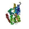
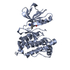
Entry Database : PDB / ID : 3a62Title Crystal structure of phosphorylated p70S6K1 Ribosomal protein S6 kinase beta-1 Keywords / / / / / / / / / / / / / / / / Function / homology Function Domain/homology Component
/ / / / / / / / / / / / / / / / / / / / / / / / / / / / / / / / / / / / / / / / / / / / / / / / / / / / / / / / / / / / / / / / / / / / / / / / / / / / / / / Biological species Homo sapiens (human)Method / / / Resolution : 2.35 Å Authors Sunami, T. / Byrne, N. / Diehl, R.E. / Funabashi, K. / Hall, D.L. / Ikuta, M. / Patel, S.B. / Shipman, J.M. / Smith, R.F. / Takahashi, I. ...Sunami, T. / Byrne, N. / Diehl, R.E. / Funabashi, K. / Hall, D.L. / Ikuta, M. / Patel, S.B. / Shipman, J.M. / Smith, R.F. / Takahashi, I. / Zugay-Murphy, J. / Iwasawa, Y. / Lumb, K.J. / Munshi, S.K. / Sharma, S. Journal : J.Biol.Chem. / Year : 2010Title : Structural basis of human p70 ribosomal S6 kinase-1 regulation by activation loop phosphorylation.Authors : Sunami, T. / Byrne, N. / Diehl, R.E. / Funabashi, K. / Hall, D.L. / Ikuta, M. / Patel, S.B. / Shipman, J.M. / Smith, R.F. / Takahashi, I. / Zugay-Murphy, J. / Iwasawa, Y. / Lumb, K.J. / Munshi, S.K. / Sharma, S. History Deposition Aug 18, 2009 Deposition site / Processing site Revision 1.0 Oct 27, 2009 Provider / Type Revision 1.1 Jul 13, 2011 Group Revision 1.2 Nov 1, 2023 Group Data collection / Database references ... Data collection / Database references / Derived calculations / Refinement description Category chem_comp_atom / chem_comp_bond ... chem_comp_atom / chem_comp_bond / database_2 / pdbx_initial_refinement_model / pdbx_struct_conn_angle / struct_conn / struct_ref_seq_dif / struct_site Item _database_2.pdbx_DOI / _database_2.pdbx_database_accession ... _database_2.pdbx_DOI / _database_2.pdbx_database_accession / _pdbx_struct_conn_angle.ptnr1_auth_comp_id / _pdbx_struct_conn_angle.ptnr1_auth_seq_id / _pdbx_struct_conn_angle.ptnr1_label_atom_id / _pdbx_struct_conn_angle.ptnr1_label_comp_id / _pdbx_struct_conn_angle.ptnr1_label_seq_id / _pdbx_struct_conn_angle.ptnr3_auth_comp_id / _pdbx_struct_conn_angle.ptnr3_auth_seq_id / _pdbx_struct_conn_angle.ptnr3_label_atom_id / _pdbx_struct_conn_angle.ptnr3_label_comp_id / _pdbx_struct_conn_angle.ptnr3_label_seq_id / _pdbx_struct_conn_angle.value / _struct_conn.pdbx_dist_value / _struct_conn.pdbx_leaving_atom_flag / _struct_conn.ptnr1_auth_comp_id / _struct_conn.ptnr1_auth_seq_id / _struct_conn.ptnr1_label_asym_id / _struct_conn.ptnr1_label_atom_id / _struct_conn.ptnr1_label_comp_id / _struct_conn.ptnr1_label_seq_id / _struct_conn.ptnr2_auth_comp_id / _struct_conn.ptnr2_auth_seq_id / _struct_conn.ptnr2_label_asym_id / _struct_conn.ptnr2_label_atom_id / _struct_conn.ptnr2_label_comp_id / _struct_conn.ptnr2_label_seq_id / _struct_ref_seq_dif.details / _struct_site.pdbx_auth_asym_id / _struct_site.pdbx_auth_comp_id / _struct_site.pdbx_auth_seq_id Revision 1.3 Oct 30, 2024 Group / Category / pdbx_modification_feature
Show all Show less
 Movie
Movie Controller


 Open data
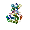
 Basic information
 Components
 Keywords
 Function and homology information
 Homo sapiens (human)
 X-RAY DIFFRACTION /
  Authors
 Citation
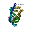
 Structure visualization
 Downloads & links
 Other downloads
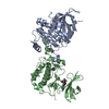

 PDBj
PDBj


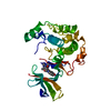
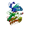
 Assembly

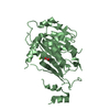
 Spodoptera frugiperda (fall armyworm)
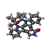
 Mass: 466.531 Da / Num. of mol.: 1 / Source method: obtained synthetically / Formula: C28H26N4O3 / Comment: anticancer, antifungal, antibiotic, alkaloid*YM
 Mass: 54.938 Da / Num. of mol.: 1 / Source method: obtained synthetically / Formula: Mn
 Mass: 18.015 Da / Num. of mol.: 37 / Source method: isolated from a natural source / Formula: H2O
 Sample preparation
 / Beamline: BL-17A / Wavelength: 1 Å
 Processing