 Movie
Movie Controller

[English] 日本語
 Yorodumi
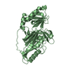
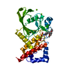
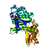
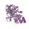
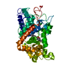
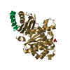
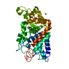
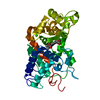
- PDB-3a55: Crystal structure of the A47Q2 mutant of pro- protein-glutaminase -
+ Open data

- Basic information
| Entry | Database: PDB / ID: 3a55 | ||||||
|---|---|---|---|---|---|---|---|
| Title | Crystal structure of the A47Q2 mutant of pro- protein-glutaminase | ||||||
 Components Components | Protein-glutaminase | ||||||
 Keywords Keywords | HYDROLASE / mutant structure like the reaction intermediate | ||||||
| Function / homology |  Function and homology information Function and homology information | ||||||
| Biological species |  Chryseobacterium proteolyticum (bacteria) Chryseobacterium proteolyticum (bacteria) | ||||||
| Method |  X-RAY DIFFRACTION / SYNCHROTRON / MOLECULAR REPLACEMENT / Resolution: 1.5 Å X-RAY DIFFRACTION / SYNCHROTRON / MOLECULAR REPLACEMENT / Resolution: 1.5 Å | ||||||
 Authors Authors | Hashizume, R. / Yamaguchi, S. / Mikami, B. | ||||||
 Citation Citation | Journal: J.Biol.Chem. / Year: 2011 Title: Crystal structures of protein glutaminase and its pro forms converted into enzyme-substrate complex Authors: Hashizume, R. / Maki, Y. / Mizutani, K. / Takahashi, N. / Matsubara, H. / Sugita, A. / Sato, K. / Yamaguchi, S. / Mikami, B. | ||||||
| History |
|
- Structure visualization
| Structure viewer | Molecule: MolmilJmol/JSmol |
|---|
- Downloads & links
-Download
| PDBx/mmCIF format | 3a55.cif.gz | 272.8 KB | Display | PDBx/mmCIF format |
|---|---|---|---|---|
| PDB format | pdb3a55.ent.gz | 221.5 KB | Display | PDB format |
| PDBx/mmJSON format | 3a55.json.gz | Tree view | PDBx/mmJSON format | |
| Others |  Other downloads Other downloads |
-Validation report
| Arichive directory | https://data.pdbj.org/pub/pdb/validation_reports/a5/3a55ftp://data.pdbj.org/pub/pdb/validation_reports/a5/3a55 | HTTPS FTP |
|---|
-Related structure data
| Related structure data | 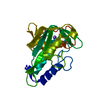 2zk9C 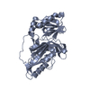 3a54C 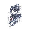 3a56SC C: citing same article ( S: Starting model for refinement |
|---|---|
| Similar structure data |


-Links
 PDBj
PDBj
- Assembly
| Deposited unit | 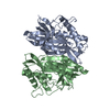
| ||||||||
|---|---|---|---|---|---|---|---|---|---|
| 1 | 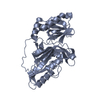
| ||||||||
| 2 | 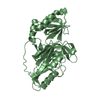
| ||||||||
| Unit cell |
|
-Components
| #1: Protein | Mass: 33611.750 Da / Num. of mol.: 2 / Mutation: A47Q Source method: isolated from a genetically manipulated source Source: (gene. exp.) Chryseobacterium proteolyticum (bacteria)Strain: DH5a / Gene: prgA / Plasmid: pET-20b / Production host: References: UniProt: Q9AQQ8, Hydrolases; Acting on carbon-nitrogen bonds, other than peptide bonds; In linear amides #2: Water | ChemComp-HOH / |  Mass: 18.015 Da / Num. of mol.: 837 / Source method: isolated from a natural source / Formula: H2O Mass: 18.015 Da / Num. of mol.: 837 / Source method: isolated from a natural source / Formula: H2OHas protein modification | Y | |
|---|
-Experimental details
-Experiment
| Experiment | Method: X-RAY DIFFRACTION / Number of used crystals: 1 |
|---|
- Sample preparation
| Crystal | Density Matthews: 2.96 Å3/Da / Density % sol: 58.41 % |
|---|---|
| Crystal grow | Temperature: 293 K / Method: vapor diffusion, hanging drop / pH: 8.7 Details: 20% PEG 3350, 0.2M sodium tartrate, pH 8.7, VAPOR DIFFUSION, HANGING DROP, temperature 293K |
-Data collection
| Diffraction | Mean temperature: 100 K |
|---|---|
| Diffraction source | Source: SYNCHROTRON / Site: SPring-8  / Beamline: BL38B1 / Wavelength: 0.8 Å / Beamline: BL38B1 / Wavelength: 0.8 Å |
| Detector | Type: RIGAKU JUPITER 210 / Detector: CCD / Date: Nov 15, 2008 |
| Radiation | Protocol: SINGLE WAVELENGTH / Monochromatic (M) / Laue (L): M / Scattering type: x-ray |
| Radiation wavelength | Wavelength: 0.8 Å / Relative weight: 1 |
| Reflection | Resolution: 1.5→50 Å / Num. obs: 119408 / % possible obs: 93.5 % / Redundancy: 4.1 % / Biso Wilson estimate: 14.99 Å2 / Rmerge(I) obs: 0.041 / Net I/σ(I): 39.27 |
| Reflection shell | Resolution: 1.5→1.53 Å / Redundancy: 3.6 % / Rmerge(I) obs: 0.332 / Num. unique all: 6016 / % possible all: 96.1 |
- Processing
| Software |
| ||||||||||||||||||||||||||||||||||||||||||||||||||||||||||||||||||||||||||||||||||||||||||||||||||||||||||||||||||||||||||||||||||||||||||||||||||||||||||||||||||||||||||
|---|---|---|---|---|---|---|---|---|---|---|---|---|---|---|---|---|---|---|---|---|---|---|---|---|---|---|---|---|---|---|---|---|---|---|---|---|---|---|---|---|---|---|---|---|---|---|---|---|---|---|---|---|---|---|---|---|---|---|---|---|---|---|---|---|---|---|---|---|---|---|---|---|---|---|---|---|---|---|---|---|---|---|---|---|---|---|---|---|---|---|---|---|---|---|---|---|---|---|---|---|---|---|---|---|---|---|---|---|---|---|---|---|---|---|---|---|---|---|---|---|---|---|---|---|---|---|---|---|---|---|---|---|---|---|---|---|---|---|---|---|---|---|---|---|---|---|---|---|---|---|---|---|---|---|---|---|---|---|---|---|---|---|---|---|---|---|---|---|---|---|---|
| Refinement | Method to determine structure: MOLECULAR REPLACEMENT Starting model: PDB ENTRY 3A56 Resolution: 1.5→50 Å / Cor.coef. Fo:Fc: 0.97 / Cor.coef. Fo:Fc free: 0.963 / SU B: 2.497 / SU ML: 0.042 / Cross valid method: THROUGHOUT / ESU R: 0.071 / ESU R Free: 0.062 / Stereochemistry target values: MAXIMUM LIKELIHOOD / Details: HYDROGENS HAVE BEEN ADDED IN THE RIDING POSITIONS
| ||||||||||||||||||||||||||||||||||||||||||||||||||||||||||||||||||||||||||||||||||||||||||||||||||||||||||||||||||||||||||||||||||||||||||||||||||||||||||||||||||||||||||
| Solvent computation | Ion probe radii: 0.8 Å / Shrinkage radii: 0.8 Å / VDW probe radii: 1.2 Å / Solvent model: MASK | ||||||||||||||||||||||||||||||||||||||||||||||||||||||||||||||||||||||||||||||||||||||||||||||||||||||||||||||||||||||||||||||||||||||||||||||||||||||||||||||||||||||||||
| Displacement parameters | Biso mean: 26.601 Å2
| ||||||||||||||||||||||||||||||||||||||||||||||||||||||||||||||||||||||||||||||||||||||||||||||||||||||||||||||||||||||||||||||||||||||||||||||||||||||||||||||||||||||||||
| Refinement step | Cycle: LAST / Resolution: 1.5→50 Å
| ||||||||||||||||||||||||||||||||||||||||||||||||||||||||||||||||||||||||||||||||||||||||||||||||||||||||||||||||||||||||||||||||||||||||||||||||||||||||||||||||||||||||||
| Refine LS restraints |
| ||||||||||||||||||||||||||||||||||||||||||||||||||||||||||||||||||||||||||||||||||||||||||||||||||||||||||||||||||||||||||||||||||||||||||||||||||||||||||||||||||||||||||
| LS refinement shell | Resolution: 1.501→1.54 Å / Total num. of bins used: 20
|
