Movie
 Controller


[English] 日本語
 Yorodumi
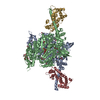
- PDB-37py: Human beta-cardiac myosin S1 motor domain with the essential ligh... -
+ Open data

- Basic information
| Entry | Database: PDB / ID: 37py | ||||||||||||
|---|---|---|---|---|---|---|---|---|---|---|---|---|---|
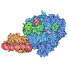
| Title | Human beta-cardiac myosin S1 motor domain with the essential light chain in the pre-powerstroke state | ||||||||||||
 Components |
| ||||||||||||
 Keywords | CONTRACTILE PROTEIN / Cardiac Myosin / Motor Domain / Essential Light Chain / Actin Binding / ADP / Phosphate | ||||||||||||
| Function / homology |  Function and homology information regulation of slow-twitch skeletal muscle fiber contraction / regulation of the force of skeletal muscle contraction / Striated Muscle Contraction / unconventional myosin complex / contractile muscle fiber / regulation of the force of heart contraction / muscle myosin complex / adult heart development / myosin filament / muscle filament sliding ...regulation of slow-twitch skeletal muscle fiber contraction / regulation of the force of skeletal muscle contraction / Striated Muscle Contraction / unconventional myosin complex / contractile muscle fiber / regulation of the force of heart contraction / muscle myosin complex / adult heart development / myosin filament / muscle filament sliding / myosin complex / myosin II complex / structural constituent of muscle / microfilament motor activity / ventricular cardiac muscle tissue morphogenesis / myofibril / cardiac muscle contraction / ATP metabolic process / skeletal muscle contraction / regulation of heart rate / stress fiber / muscle contraction / bioluminescence / sarcomere / generation of precursor metabolites and energy / striated muscle contraction / Z disc / actin filament binding / calcium ion binding / ATP binding / cytoplasm Similarity search - Function | ||||||||||||
| Biological species |  Homo sapiens (human)  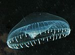 Aequorea victoria (jellyfish)   Mus musculus (house mouse) | ||||||||||||
| Method | ELECTRON MICROSCOPY / single particle reconstruction / cryo EM / Resolution: 3.2 Å | ||||||||||||
 Authors | Somavarapu, A.K. / Gautam, R. / Craig, R. / Padron, R. | ||||||||||||
| Funding support |  United States, 3items
| ||||||||||||
 Citation | Journal: bioRxiv / Year: 2026 Title: The dilated cardiomyopathy E525K mutation stabilizes the cardiac myosin interacting-heads motif while activating the isolated motor domain. Authors: Ruchi Gautam / Arun Kumar Somavarapu / Kalen Z Robeson / Matthew C Childers / Michael Regnier / Jinghua Ge / Christopher M Yengo / Roger Craig / Raúl Padrón / Abstract: Mutations in β-cardiac myosin are a common cause of inherited cardiomyopathies. The dilated cardiomyopathy E525K mutation alters both thick filament regulation and motor activity, yet the structural ...Mutations in β-cardiac myosin are a common cause of inherited cardiomyopathies. The dilated cardiomyopathy E525K mutation alters both thick filament regulation and motor activity, yet the structural basis for these effects has remained unclear. Here, we combined cryo-EM and MD simulations to determine how E525K affects the conformational landscape of cardiac myosin in both its autoinhibited interacting-heads motif (IHM) and isolated myosin head states. E525K-mutated myosin exclusively adopted a single S2 conformation in IHM, in contrast to the conformational heterogeneity observed in wild-type myosin. Structural and electrostatic analyses revealed that the mutation increased positive charge density at the blocked-head (BH) - S2 (subfragment-2) interface, strengthening interactions with negatively charged residues within S2. 3D variability analysis and MD simulations of the E525K IHM demonstrated reduced S2 mobility, consistent with strengthened BH - S2 interactions, and decreased conformational flexibility, providing a structural mechanism for stabilization of the IHM state. To investigate the isolated myosin head, we determined cryo-EM structures of wild-type and E525K subfragment-1 (S1). The mutation induced local conformational rearrangements in the activation-loop and Loop 3 region, the SH3-like domain, and the essential light chain of S1, revealing structural changes that support the enhanced motor activity previously reported for isolated E525K myosin motors. These findings show that E525K stabilizes the autoinhibited IHM through electrostatic stabilization of the BH - S2 interface while inducing structural changes consistent with activation of the isolated motor domain, providing structural insights into how a single DCM mutation differentially regulates cardiac myosin S1 and IHM structure and function. | ||||||||||||
| History |
|
- Structure visualization
| Structure viewer | Molecule: MolmilJmol/JSmol |
|---|
- Downloads & links
-Download
| PDBx/mmCIF format | 37py.cif.gz | 205.4 KB | Display | PDBx/mmCIF format |
|---|---|---|---|---|
| PDB format | pdb37py.ent.gz | 153.6 KB | Display | PDB format |
| PDBx/mmJSON format | 37py.json.gz | Tree view | PDBx/mmJSON format | |
| Others |  Other downloads |
-Validation report
| Arichive directory | https://data.pdbj.org/pub/pdb/validation_reports/7p/37pyftp://data.pdbj.org/pub/pdb/validation_reports/7p/37py | HTTPS FTP |
|---|
-Related structure data
| Related structure data |  78401MC  37oaC  37qcC M: map data used to model this data C: citing same article ( |
|---|---|
| Similar structure data |


-Links
 PDBj

- Assembly
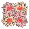
| Deposited unit | 
|
|---|---|
| 1 |
|
-Components
| #1: Protein | Mass: 126485.500 Da / Num. of mol.: 1 Source method: isolated from a genetically manipulated source Source: (gene. exp.) Homo sapiens (human), (gene. exp.) Aequorea victoria (jellyfish)Gene: MYH7, MYHCB, GFP / Production host: Mus musculus (house mouse) / References: UniProt: P12883, UniProt: P42212 |
|---|---|
| #2: Protein | Mass: 20620.490 Da / Num. of mol.: 1 / Source method: isolated from a natural source / Source: (natural) Mus musculus (house mouse) / References: UniProt: P05977 |
| #3: Chemical | ChemComp-ADP / 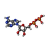  Mass: 427.201 Da / Num. of mol.: 1 / Source method: isolated from a natural source / Formula: C10H15N5O10P2 / Feature type: SUBJECT OF INVESTIGATION / Comment: ADP, energy-carrying molecule*YM |
| #4: Chemical | ChemComp-PO4 / 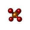  Mass: 94.971 Da / Num. of mol.: 1 / Source method: obtained synthetically / Formula: PO4 / Feature type: SUBJECT OF INVESTIGATION |
| #5: Chemical | ChemComp-MG /   Mass: 24.305 Da / Num. of mol.: 1 / Source method: obtained synthetically / Formula: Mg / Feature type: SUBJECT OF INVESTIGATION |
| Has ligand of interest | Y |
| Has protein modification | N |
-Experimental details
-Experiment
| Experiment | Method: ELECTRON MICROSCOPY |
|---|---|
| EM experiment | Aggregation state: PARTICLE / 3D reconstruction method: single particle reconstruction |
- Sample preparation
| Component |
| ||||||||||||||||||||||||
|---|---|---|---|---|---|---|---|---|---|---|---|---|---|---|---|---|---|---|---|---|---|---|---|---|---|
| Molecular weight | Value: 0.11652168 MDa / Experimental value: NO | ||||||||||||||||||||||||
| Source (natural) |
| ||||||||||||||||||||||||
| Source (recombinant) | Organism: Mus musculus (house mouse) | ||||||||||||||||||||||||
| Buffer solution | pH: 7.4 | ||||||||||||||||||||||||
| Specimen | Embedding applied: NO / Shadowing applied: NO / Staining applied: NO / Vitrification applied: YES | ||||||||||||||||||||||||
| Specimen support | Grid material: GOLD / Grid type: UltrAuFoil R1.2/1.3 | ||||||||||||||||||||||||
| Vitrification | Instrument: FEI VITROBOT MARK IV / Cryogen name: ETHANE |
- Electron microscopy imaging
| Experimental equipment | 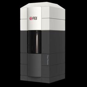 Model: Titan Krios / Image courtesy: FEI Company |
|---|---|
| Microscopy | Model: TFS KRIOS |
| Electron gun | Electron source:  FIELD EMISSION GUN / Accelerating voltage: 300 kV / Illumination mode: FLOOD BEAM |
| Electron lens | Mode: BRIGHT FIELD / Nominal defocus max: 2000 nm / Nominal defocus min: 1000 nm |
| Specimen holder | Specimen holder model: FEI TITAN KRIOS AUTOGRID HOLDER |
| Image recording | Electron dose: 50.5 e/Å2 / Film or detector model: GATAN K3 (6k x 4k) |
- Processing
| EM software |
| ||||||||||||||||||||||||||||||||||||||||
|---|---|---|---|---|---|---|---|---|---|---|---|---|---|---|---|---|---|---|---|---|---|---|---|---|---|---|---|---|---|---|---|---|---|---|---|---|---|---|---|---|---|
| CTF correction | Type: PHASE FLIPPING AND AMPLITUDE CORRECTION | ||||||||||||||||||||||||||||||||||||||||
| 3D reconstruction | Resolution: 3.2 Å / Resolution method: FSC 0.143 CUT-OFF / Num. of particles: 90877 / Symmetry type: POINT | ||||||||||||||||||||||||||||||||||||||||
| Atomic model building | Protocol: FLEXIBLE FIT / Space: REAL | ||||||||||||||||||||||||||||||||||||||||
| Atomic model building | PDB-ID: 5N69 Accession code: 5N69 / Source name: PDB / Type: experimental model | ||||||||||||||||||||||||||||||||||||||||
| Refinement | Highest resolution: 3.2 Å Stereochemistry target values: REAL-SPACE (WEIGHTED MAP SUM AT ATOM CENTERS) | ||||||||||||||||||||||||||||||||||||||||
| Refine LS restraints |
|