[English] 日本語
Yorodumi
- EMDB-78401: Human beta-cardiac myosin S1 motor domain with the essential ligh... -

+
Open data


ID or keywords:

Loading...

-
Basic information

Entry
Database: EMDB / ID: EMD-78401
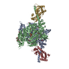
TitleHuman beta-cardiac myosin S1 motor domain with the essential light chain in the pre-powerstroke state
Map data
Sample
  • Complex: Human beta-cardiac myosin S1 motor domain with the essential light chain in the pre-powerstroke state
    • Complex: Myosin-7
      • Protein or peptide: Myosin-7,Green fluorescent protein
    • Complex: Myosin light chain 1/3
      • Protein or peptide: Myosin light chain 1/3, skeletal muscle isoform
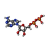
  • Ligand: ADENOSINE-5'-DIPHOSPHATE
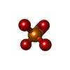
  • Ligand: PHOSPHATE ION
  • Ligand: MAGNESIUM ION
KeywordsCardiac Myosin / Motor Domain / Essential Light Chain / Contractile Protein / Actin Binding / ADP / Phosphate
Function / homology
Function and homology information


regulation of slow-twitch skeletal muscle fiber contraction / regulation of the force of skeletal muscle contraction / Striated Muscle Contraction / unconventional myosin complex / contractile muscle fiber / regulation of the force of heart contraction / muscle myosin complex / adult heart development / myosin filament / muscle filament sliding ...regulation of slow-twitch skeletal muscle fiber contraction / regulation of the force of skeletal muscle contraction / Striated Muscle Contraction / unconventional myosin complex / contractile muscle fiber / regulation of the force of heart contraction / muscle myosin complex / adult heart development / myosin filament / muscle filament sliding / myosin complex / myosin II complex / structural constituent of muscle / microfilament motor activity / ventricular cardiac muscle tissue morphogenesis / myofibril / cardiac muscle contraction / ATP metabolic process / skeletal muscle contraction / regulation of heart rate / stress fiber / muscle contraction / bioluminescence / sarcomere / generation of precursor metabolites and energy / striated muscle contraction / Z disc / actin filament binding / calcium ion binding / ATP binding / cytoplasm
Similarity search - Function
DNA repair protein XRCC4-like, C-terminal / Myosin tail / Myosin tail / Myosin N-terminal SH3-like domain / Myosin S1 fragment, N-terminal / Myosin, N-terminal, SH3-like / Myosin N-terminal SH3-like domain profile. / Short calmodulin-binding motif containing conserved Ile and Gln residues. / IQ motif, EF-hand binding site / Myosin motor domain profile. ...DNA repair protein XRCC4-like, C-terminal / Myosin tail / Myosin tail / Myosin N-terminal SH3-like domain / Myosin S1 fragment, N-terminal / Myosin, N-terminal, SH3-like / Myosin N-terminal SH3-like domain profile. / Short calmodulin-binding motif containing conserved Ile and Gln residues. / IQ motif, EF-hand binding site / Myosin motor domain profile. / Myosin head, motor domain / Myosin head (motor domain) / Myosin. Large ATPases. / IQ motif profile. / Kinesin motor domain superfamily / : / Green fluorescent protein, GFP / Green fluorescent protein-related / Green fluorescent protein / Green fluorescent protein / EF-hand calcium-binding domain profile. / EF-hand domain / EF-hand domain pair / P-loop containing nucleoside triphosphate hydrolase
Similarity search - Domain/homology
Myosin light chain 1/3, skeletal muscle isoform / Myosin-7 / Green fluorescent protein
Similarity search - Component
Biological speciesHomo sapiens (human) / Mus musculus (house mouse) / Aequorea victoria (jellyfish)
Methodsingle particle reconstruction / cryo EM / Resolution: 3.2 Å
AuthorsSomavarapu AK / Gautam R / Craig R / Padron R
Funding support United States, 3 items
OrganizationGrant numberCountry
National Institutes of Health/National Heart, Lung, and Blood Institute (NIH/NHLBI)HL164560 United States
National Institutes of Health/National Institute of Arthritis and Musculoskeletal and Skin Diseases (NIH/NIAMS)AR081941 United States
American Heart Association26CDA1600784 United States
CitationJournal: bioRxiv / Year: 2026
Title: The dilated cardiomyopathy E525K mutation stabilizes the cardiac myosin interacting-heads motif while activating the isolated motor domain.
Authors: Ruchi Gautam / Arun Kumar Somavarapu / Kalen Z Robeson / Matthew C Childers / Michael Regnier / Jinghua Ge / Christopher M Yengo / Roger Craig / Raúl Padrón /
Abstract: Mutations in β-cardiac myosin are a common cause of inherited cardiomyopathies. The dilated cardiomyopathy E525K mutation alters both thick filament regulation and motor activity, yet the structural ...Mutations in β-cardiac myosin are a common cause of inherited cardiomyopathies. The dilated cardiomyopathy E525K mutation alters both thick filament regulation and motor activity, yet the structural basis for these effects has remained unclear. Here, we combined cryo-EM and MD simulations to determine how E525K affects the conformational landscape of cardiac myosin in both its autoinhibited interacting-heads motif (IHM) and isolated myosin head states. E525K-mutated myosin exclusively adopted a single S2 conformation in IHM, in contrast to the conformational heterogeneity observed in wild-type myosin. Structural and electrostatic analyses revealed that the mutation increased positive charge density at the blocked-head (BH) - S2 (subfragment-2) interface, strengthening interactions with negatively charged residues within S2. 3D variability analysis and MD simulations of the E525K IHM demonstrated reduced S2 mobility, consistent with strengthened BH - S2 interactions, and decreased conformational flexibility, providing a structural mechanism for stabilization of the IHM state. To investigate the isolated myosin head, we determined cryo-EM structures of wild-type and E525K subfragment-1 (S1). The mutation induced local conformational rearrangements in the activation-loop and Loop 3 region, the SH3-like domain, and the essential light chain of S1, revealing structural changes that support the enhanced motor activity previously reported for isolated E525K myosin motors. These findings show that E525K stabilizes the autoinhibited IHM through electrostatic stabilization of the BH - S2 interface while inducing structural changes consistent with activation of the isolated motor domain, providing structural insights into how a single DCM mutation differentially regulates cardiac myosin S1 and IHM structure and function.
History
DepositionJul 31, 2026-
Header (metadata) releaseNov 4, 2026-
Map releaseNov 4, 2026-
UpdateNov 4, 2026-
Current statusNov 4, 2026Processing site: RCSB / Status: Released

-
Structure visualization

Supplemental images

Downloads & links

-
Map

FileDownload / File: emd_78401.map.gz / Format: CCP4 / Size: 64 MB / Type: IMAGE STORED AS FLOATING POINT NUMBER (4 BYTES)
Projections & slices

Image control

Size
Brightness
Contrast
Others
AxesZ (Sec.)Y (Row.)X (Col.)
0.83 Å/pix.
x 256 pix.
= 212.48 Å
0.83 Å/pix.
x 256 pix.
= 212.48 Å
0.83 Å/pix.
x 256 pix.
= 212.48 Å

Surface

Projections

Slices (1/3)

Slices (1/2)

Slices (2/3)

Images are generated by Spider.

Voxel sizeX=Y=Z: 0.83 Å
Density
Contour LevelBy AUTHOR: 0.05
Minimum - Maximum-0.67630184 - 1.0022852
Average (Standard dev.)0.00088560587 (±0.021086913)
SymmetrySpace group: 1
Details

EMDB XML:

Map geometry
Axis orderXYZ
Origin000
Dimensions256256256
Spacing256256256
CellA=B=C: 212.48 Å
α=β=γ: 90.0 °

-
Supplemental data

-
Mask #1

Fileemd_78401_msk_1.map
Projections & Slices
AxesZYX

Projections

Slices (1/2)
Density Histograms

-
Half map: #2

Fileemd_78401_half_map_1.map
Projections & Slices
AxesZYX

Projections

Slices (1/2)
Density Histograms

-
Half map: #1

Fileemd_78401_half_map_2.map
Projections & Slices
AxesZYX

Projections

Slices (1/2)
Density Histograms

-
Sample components

-
Entire : Human beta-cardiac myosin S1 motor domain with the essential ligh...

EntireName: Human beta-cardiac myosin S1 motor domain with the essential light chain in the pre-powerstroke state
Components
  • Complex: Human beta-cardiac myosin S1 motor domain with the essential light chain in the pre-powerstroke state
    • Complex: Myosin-7
      • Protein or peptide: Myosin-7,Green fluorescent protein
    • Complex: Myosin light chain 1/3
      • Protein or peptide: Myosin light chain 1/3, skeletal muscle isoform
  • Ligand: ADENOSINE-5'-DIPHOSPHATE
  • Ligand: PHOSPHATE ION
  • Ligand: MAGNESIUM ION

-
Supramolecule #1: Human beta-cardiac myosin S1 motor domain with the essential ligh...

SupramoleculeName: Human beta-cardiac myosin S1 motor domain with the essential light chain in the pre-powerstroke state
type: complex / ID: 1 / Parent: 0 / Macromolecule list: #1-#2
Molecular weightTheoretical: 116.52168 KDa

-
Supramolecule #2: Myosin-7

SupramoleculeName: Myosin-7 / type: complex / ID: 2 / Parent: 1 / Macromolecule list: #1
Source (natural)Organism: Homo sapiens (human)

-
Supramolecule #3: Myosin light chain 1/3

SupramoleculeName: Myosin light chain 1/3 / type: complex / ID: 3 / Parent: 1 / Macromolecule list: #2
Source (natural)Organism: Mus musculus (house mouse)

-
Macromolecule #1: Myosin-7,Green fluorescent protein

MacromoleculeName: Myosin-7,Green fluorescent protein / type: protein_or_peptide / ID: 1 / Number of copies: 1 / Enantiomer: LEVO
Source (natural)Organism: Aequorea victoria (jellyfish)
Molecular weightTheoretical: 126.4855 KDa
Recombinant expressionOrganism: Mus musculus (house mouse)
SequenceString: MGDSEMAVFG AAAPYLRKSE KERLEAQTRP FDLKKDVFVP DDKQEFVKAK IVSREGGKVT AETEYGKTVT VKEDQVMQQN PPKFDKIED MAMLTFLHEP AVLYNLKDRY GSWMIYTYSG LFCVTVNPYK WLPVYTPEVV AAYRGKKRSE APPHIFSISD N AYQYMLTD ...String:
MGDSEMAVFG AAAPYLRKSE KERLEAQTRP FDLKKDVFVP DDKQEFVKAK IVSREGGKVT AETEYGKTVT VKEDQVMQQN PPKFDKIED MAMLTFLHEP AVLYNLKDRY GSWMIYTYSG LFCVTVNPYK WLPVYTPEVV AAYRGKKRSE APPHIFSISD N AYQYMLTD RENQSILITG ESGAGKTVNT KRVIQYFAVI AAIGDRSKKD QSPGKGTLED QIIQANPALE AFGNAKTVRN DN SSRFGKF IRIHFGATGK LASADIETYL LEKSRVIFQL KAERDYHIFY QILSNKKPEL LDMLLITNNP YDYAFISQGE TTV ASIDDA EELMATDNAF DVLGFTSEEK NSMYKLTGAI MHFGNMKFKL KQREEQAEPD GTEEADKSAY LMGLNSADLL KGLC HPRVK VGNEYVTKGQ NVQQVIYATG ALAKAVYERM FNWMVTRINA TLETKQPRQY FIGVLDIAGF EIFDFNSFEQ LCINF TNEK LQQFFNHHMF VLEQEEYKKE GIEWTFIDFG MDLQACIDLI EKPMGIMSIL EEECMFPKAT DMTFKAKLFD NHLGKS ANF QKPRNIKGKP EAHFSLIHYA GIVDYNIIGW LQKNKDPLNE TVVGLYQKSS LKLLSTLFAN YAGADAPIEK GKGKAKK GS SFQTVSALHR ENLNKLMTNL RSTHPHFVRC IIPNETKSPG VMDNPLVMHQ LRCNGVLEGI RICRKGFPNR ILYGDFRQ R YRILNPAAIP EGQFIDSRKG AEKLLSSLDI DHNQYKFGHT KVFFKAGLLG LLEEMRDERL SRIITRIQAQ SRGVLARME YKKLLERRDS LLVIQWNIRA FMGVKNWPWM KLYFKIKPLL KSGSGKLGVS KGEELFTGVV PILVELDGDV NGHKFSVSGE GEGDATYGK LTLKFICTTG KLPVPWPTLV TTLTYGVQCF SRYPDHMKQH DFFKSAMPEG YVQERTIFFK DDGNYKTRAE V KFEGDTLV NRIELKGIDF KEDGNILGHK LEYNYNSHNV YIMADKQKNG IKVNFKIRHN IEDGSVQLAD HYQQNTPIGD GP VLLPDNH YLSTQSALSK DPNEKRDHMV LLEFVTAAGI TLGMDELYKG LNDIFEAQKI EWHEDYKDDD DK

UniProtKB: Myosin-7, Green fluorescent protein

-
Macromolecule #2: Myosin light chain 1/3, skeletal muscle isoform

MacromoleculeName: Myosin light chain 1/3, skeletal muscle isoform / type: protein_or_peptide / ID: 2 / Number of copies: 1 / Enantiomer: LEVO
Source (natural)Organism: Mus musculus (house mouse)
Molecular weightTheoretical: 20.62049 KDa
SequenceString:
MAPKKDVKKP AAAPAPAPAP APAPAKPKEE KIDLSAIKIE FSKEQQEDFK EAFLLFDRTG ECKITLSQVG DVLRALGTNP TNAEVKKVL GNPSNEEMNA KKIEFEQFLP MMQAISNNKD QGGYEDFVEG LRVFDKEGNG TVMGAELRHV LATLGEKMKE E EVEALLAG QEDSNGCINY EAFVKHIMSV

UniProtKB: Myosin light chain 1/3, skeletal muscle isoform

-
Macromolecule #3: ADENOSINE-5'-DIPHOSPHATE

MacromoleculeName: ADENOSINE-5'-DIPHOSPHATE / type: ligand / ID: 3 / Number of copies: 1 / Formula: ADP
Molecular weightTheoretical: 427.201 Da
Chemical component information

ChemComp-ADP:
ADENOSINE-5'-DIPHOSPHATE / ADP, energy-carrying molecule*YM

-
Macromolecule #4: PHOSPHATE ION

MacromoleculeName: PHOSPHATE ION / type: ligand / ID: 4 / Number of copies: 1 / Formula: PO4
Molecular weightTheoretical: 94.971 Da
Chemical component information

ChemComp-PO4:
PHOSPHATE ION

-
Macromolecule #5: MAGNESIUM ION

MacromoleculeName: MAGNESIUM ION / type: ligand / ID: 5 / Number of copies: 1 / Formula: MG
Molecular weightTheoretical: 24.305 Da

-
Experimental details

-
Structure determination

Methodcryo EM
Processingsingle particle reconstruction
Aggregation stateparticle

-
Sample preparation

BufferpH: 7.4
GridModel: UltrAuFoil R1.2/1.3 / Material: GOLD / Pretreatment - Type: GLOW DISCHARGE
VitrificationCryogen name: ETHANE / Instrument: FEI VITROBOT MARK IV

-
Electron microscopy

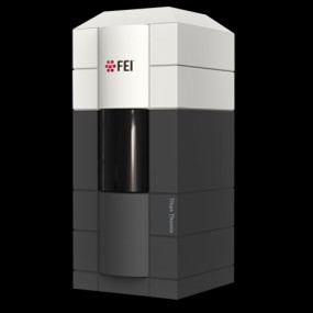
MicroscopeTFS KRIOS
Image recordingFilm or detector model: GATAN K3 (6k x 4k) / Average electron dose: 50.5 e/Å2
Electron beamAcceleration voltage: 300 kV / Electron source: FIELD EMISSION GUN
Electron opticsIllumination mode: FLOOD BEAM / Imaging mode: BRIGHT FIELD / Nominal defocus max: 2.0 µm / Nominal defocus min: 1.0 µm
Sample stageSpecimen holder model: FEI TITAN KRIOS AUTOGRID HOLDER
Experimental equipment
Model: Titan Krios / Image courtesy: FEI Company

+
Image processing

CTF correctionSoftware - Name: cryoSPARC / Type: PHASE FLIPPING AND AMPLITUDE CORRECTION
Startup modelType of model: NONE
Final reconstructionResolution.type: BY AUTHOR / Resolution: 3.2 Å / Resolution method: FSC 0.143 CUT-OFF / Software - Name: cryoSPARC / Number images used: 90877
Initial angle assignmentType: MAXIMUM LIKELIHOOD / Software - Name: cryoSPARC
Final angle assignmentType: MAXIMUM LIKELIHOOD / Software - Name: cryoSPARC
FSC plot (resolution estimation)

-
Atomic model buiding 1

Initial modelPDB ID:

Chain - Source name: PDB / Chain - Initial model type: experimental model
SoftwareName: UCSF ChimeraX
RefinementSpace: REAL / Protocol: FLEXIBLE FIT
Output model

PDB-37py:
Human beta-cardiac myosin S1 motor domain with the essential light chain in the pre-powerstroke state

+
About Yorodumi

-
News

-
Feb 9, 2022. New format data for meta-information of EMDB entries

New format data for meta-information of EMDB entries

  • Version 3 of the EMDB header file is now the official format.
  • The previous official version 1.9 will be removed from the archive.

Related info.:EMDB header

External links:wwPDB to switch to version 3 of the EMDB data model

-
Aug 12, 2020. Covid-19 info

Covid-19 info

URL: https://pdbj.org/emnavi/covid19.php

New page: Covid-19 featured information page in EM Navigator.

Related info.:Covid-19 info / Mar 5, 2020. Novel coronavirus structure data

+
Mar 5, 2020. Novel coronavirus structure data

Novel coronavirus structure data

Related info.:Yorodumi Speices / Aug 12, 2020. Covid-19 info

External links:COVID-19 featured content - PDBj / Molecule of the Month (242):Coronavirus Proteases

+
Jan 31, 2019. EMDB accession codes are about to change! (news from PDBe EMDB page)

EMDB accession codes are about to change! (news from PDBe EMDB page)

  • The allocation of 4 digits for EMDB accession codes will soon come to an end. Whilst these codes will remain in use, new EMDB accession codes will include an additional digit and will expand incrementally as the available range of codes is exhausted. The current 4-digit format prefixed with “EMD-” (i.e. EMD-XXXX) will advance to a 5-digit format (i.e. EMD-XXXXX), and so on. It is currently estimated that the 4-digit codes will be depleted around Spring 2019, at which point the 5-digit format will come into force.
  • The EM Navigator/Yorodumi systems omit the EMD- prefix.

Related info.:Q: What is EMD? / ID/Accession-code notation in Yorodumi/EM Navigator

External links:EMDB Accession Codes are Changing Soon! / Contact to PDBj

+
Jul 12, 2017. Major update of PDB

Major update of PDB

  • wwPDB released updated PDB data conforming to the new PDBx/mmCIF dictionary.
  • This is a major update changing the version number from 4 to 5, and with Remediation, in which all the entries are updated.
  • In this update, many items about electron microscopy experimental information are reorganized (e.g. em_software).
  • Now, EM Navigator and Yorodumi are based on the updated data.

External links:wwPDB Remediation / Enriched Model Files Conforming to OneDep Data Standards Now Available in the PDB FTP Archive

-
Yorodumi

Thousand views of thousand structures

  • Yorodumi is a browser for structure data from EMDB, PDB, SASBDB, etc.
  • This page is also the successor to EM Navigator detail page, and also detail information page/front-end page for Omokage search.
  • The word "yorodu" (or yorozu) is an old Japanese word meaning "ten thousand". "mi" (miru) is to see.

Related info.:EMDB / PDB / SASBDB / Comparison of 3 databanks / Yorodumi Search / Aug 31, 2016. New EM Navigator & Yorodumi / Yorodumi Papers / Jmol/JSmol / Function and homology information / Changes in new EM Navigator and Yorodumi

Read more