 Movie
Movie Controller


+ Open data

- Basic information
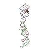
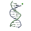
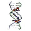
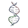
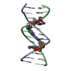
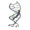
| Entry | Database: PDB / ID: 354d | ||||||
|---|---|---|---|---|---|---|---|
| Title | Structure of loop E FROM E. coli 5S RRNA | ||||||
 Components Components |
| ||||||
 Keywords Keywords | RNA / U-RNA / DOUBLE HELIX / INTERNAL LOOP / OVERHANGING BASE / MISMATCHED | ||||||
| Function / homology | RNA / RNA (> 10) Function and homology information Function and homology information | ||||||
| Biological species |  | ||||||
| Method |  X-RAY DIFFRACTION / SYNCHROTRON / MOLECULAR REPLACEMENT / Resolution: 1.5 Å X-RAY DIFFRACTION / SYNCHROTRON / MOLECULAR REPLACEMENT / Resolution: 1.5 Å | ||||||
 Authors Authors | Correll, C.C. / Freeborn, B. / Moore, P.B. / Steitz, T.A. | ||||||
 Citation Citation | Journal: Cell(Cambridge,Mass.) / Year: 1997 Title: Metals, motifs, and recognition in the crystal structure of a 5S rRNA domain. Authors: Correll, C.C. / Freeborn, B. / Moore, P.B. / Steitz, T.A. #1: Journal: J.Biomol.Struct.Dyn. / Year: 1997Title: Use of Chemically Modified Nucleotides to Determine a 62-Nucleotide RNA Crystal Structure: A Survey of Phosphorothioates, Br, Pt, and Hg Authors: Correll, C.C. / Freeborn, B. / Moore, P.B. / Steitz, T.A. #2: Journal: To be PublishedTitle: The Solution Structure of the Loop E/Loop D Region of E. coli 5S rRNA Authors: Dallas, A. / Moore, P.B. | ||||||
| History |
|
- Structure visualization
| Structure viewer | Molecule: MolmilJmol/JSmol |
|---|
- Downloads & links
-Download
| PDBx/mmCIF format | 354d.cif.gz | 29.6 KB | Display | PDBx/mmCIF format |
|---|---|---|---|---|
| PDB format | pdb354d.ent.gz | 18.8 KB | Display | PDB format |
| PDBx/mmJSON format | 354d.json.gz | Tree view | PDBx/mmJSON format | |
| Others |  Other downloads Other downloads |
-Validation report
| Arichive directory | https://data.pdbj.org/pub/pdb/validation_reports/54/354dftp://data.pdbj.org/pub/pdb/validation_reports/54/354d | HTTPS FTP |
|---|
-Related structure data


-Links
 PDBj
PDBj


- Assembly
| Deposited unit | 
| ||||||||
|---|---|---|---|---|---|---|---|---|---|
| 1 |
| ||||||||
| Unit cell |
|
-Components
| #1: RNA chain | Mass: 3868.344 Da / Num. of mol.: 1 Source method: isolated from a genetically manipulated source Source: (gene. exp.) | ||
|---|---|---|---|
| #2: RNA chain | Mass: 3930.489 Da / Num. of mol.: 1 Source method: isolated from a genetically manipulated source Source: (gene. exp.) Keywords: MUTANT: G107 IS MUTATED TO C | ||
| #3: Chemical | ChemComp-MG /   Mass: 24.305 Da / Num. of mol.: 5 / Source method: obtained synthetically / Formula: Mg Mass: 24.305 Da / Num. of mol.: 5 / Source method: obtained synthetically / Formula: Mg#4: Water | ChemComp-HOH / |  Mass: 18.015 Da / Num. of mol.: 124 / Source method: isolated from a natural source / Formula: H2O Mass: 18.015 Da / Num. of mol.: 124 / Source method: isolated from a natural source / Formula: H2O |
-Experimental details
-Experiment
| Experiment | Method: X-RAY DIFFRACTION / Number of used crystals: 1 |
|---|
- Sample preparation
| Crystal | Density Matthews: 2.72 Å3/Da / Density % sol: 54.82 % | ||||||||||||||||||||||||||||||||||||||||||
|---|---|---|---|---|---|---|---|---|---|---|---|---|---|---|---|---|---|---|---|---|---|---|---|---|---|---|---|---|---|---|---|---|---|---|---|---|---|---|---|---|---|---|---|
| Crystal grow | Method: vapor diffusion / pH: 6 / Details: pH 6.00, VAPOR DIFFUSION | ||||||||||||||||||||||||||||||||||||||||||
| Crystal grow | *PLUS Temperature: 20 ℃ / pH: 6 | ||||||||||||||||||||||||||||||||||||||||||
| Components of the solutions | *PLUS
|
-Data collection
| Diffraction | Mean temperature: 100 K |
|---|---|
| Diffraction source | Source: SYNCHROTRON / Site: CHESS  / Beamline: A1 / Beamline: A1 |
| Detector | Type: PRINCETON 2K / Detector: CCD / Date: Sep 9, 1996 / Details: SILICON 111 BENDING MIRROR |
| Radiation | Monochromatic (M) / Laue (L): M / Scattering type: x-ray |
| Radiation wavelength | Relative weight: 1 |
| Reflection | Resolution: 1.5→20 Å / Num. obs: 12306 / % possible obs: 90.7 % / Observed criterion σ(F): -3 / Observed criterion σ(I): -3 / Redundancy: 4.6 % / Biso Wilson estimate: 19.4 Å2 / Rmerge(I) obs: 0.055 / Rsym value: 0.055 / Net I/σ(I): 7.1 |
| Reflection shell | Resolution: 1.5→1.53 Å / Rmerge(I) obs: 0.48 / Mean I/σ(I) obs: 2 / Rsym value: 0.48 / % possible all: 54 |
| Reflection | *PLUS Highest resolution: 1.5 Å / Lowest resolution: 20 Å / % possible obs: 90.7 % / Observed criterion σ(F): -3 / Observed criterion σ(I): -3 / Redundancy: 4.6 % |
| Reflection shell | *PLUS Highest resolution: 1.5 Å / Lowest resolution: 1.53 Å / Mean I/σ(I) obs: 2 |
- Processing
| Software |
| ||||||||||||||||||||||||||||||||||||||||||||||||||||||||||||||||||||||||||||||||
|---|---|---|---|---|---|---|---|---|---|---|---|---|---|---|---|---|---|---|---|---|---|---|---|---|---|---|---|---|---|---|---|---|---|---|---|---|---|---|---|---|---|---|---|---|---|---|---|---|---|---|---|---|---|---|---|---|---|---|---|---|---|---|---|---|---|---|---|---|---|---|---|---|---|---|---|---|---|---|---|---|---|
| Refinement | Method to determine structure: MOLECULAR REPLACEMENT / Resolution: 1.5→20 Å / Rfactor Rfree error: 0.007 / Data cutoff high absF: 4170208 / Data cutoff low absF: 0 / Cross valid method: THROUGHOUT / σ(F): 0 Details: THERE IS A DIASTEREOMERIC MIXTURE OF RP AND SP PHOSPHOROTHIOATE AT G106, BOTH FORMS ARE 0.5 OCCUPANCY AS ALTERNATE CONFORMATIONS. WATER MEDIATED BASE-PAIRS: N3 U 74 - HOH 329 - N2 G 102 N1 G ...Details: THERE IS A DIASTEREOMERIC MIXTURE OF RP AND SP PHOSPHOROTHIOATE AT G106, BOTH FORMS ARE 0.5 OCCUPANCY AS ALTERNATE CONFORMATIONS. WATER MEDIATED BASE-PAIRS: N3 U 74 - HOH 329 - N2 G 102 N1 G 75 - HOH 324 - N1 A 101 O6 G 76 - HOH 330 - N7 G 100
| ||||||||||||||||||||||||||||||||||||||||||||||||||||||||||||||||||||||||||||||||
| Displacement parameters | Biso mean: 24.2 Å2
| ||||||||||||||||||||||||||||||||||||||||||||||||||||||||||||||||||||||||||||||||
| Refine analyze |
| ||||||||||||||||||||||||||||||||||||||||||||||||||||||||||||||||||||||||||||||||
| Refinement step | Cycle: LAST / Resolution: 1.5→20 Å
| ||||||||||||||||||||||||||||||||||||||||||||||||||||||||||||||||||||||||||||||||
| Refine LS restraints |
| ||||||||||||||||||||||||||||||||||||||||||||||||||||||||||||||||||||||||||||||||
| LS refinement shell | Resolution: 1.5→1.58 Å / Rfactor Rfree error: 0.03 / Total num. of bins used: 7
| ||||||||||||||||||||||||||||||||||||||||||||||||||||||||||||||||||||||||||||||||
| Xplor file |
| ||||||||||||||||||||||||||||||||||||||||||||||||||||||||||||||||||||||||||||||||
| Software | *PLUS Name: CNS / Version: 0.1 / Classification: refinement | ||||||||||||||||||||||||||||||||||||||||||||||||||||||||||||||||||||||||||||||||
| Refinement | *PLUS Highest resolution: 1.5 Å / Lowest resolution: 20 Å / σ(F): 0 | ||||||||||||||||||||||||||||||||||||||||||||||||||||||||||||||||||||||||||||||||
| Solvent computation | *PLUS | ||||||||||||||||||||||||||||||||||||||||||||||||||||||||||||||||||||||||||||||||
| Displacement parameters | *PLUS Biso mean: 24.2 Å2 | ||||||||||||||||||||||||||||||||||||||||||||||||||||||||||||||||||||||||||||||||
| Refine LS restraints | *PLUS
| ||||||||||||||||||||||||||||||||||||||||||||||||||||||||||||||||||||||||||||||||
| LS refinement shell | *PLUS Highest resolution: 1.5 Å / % reflection Rfree: 5 % |
