Movie
 Controller


+ Open data

- Basic information
| Entry | Database: PDB / ID: 34oa | |||||||||||||||
|---|---|---|---|---|---|---|---|---|---|---|---|---|---|---|---|---|
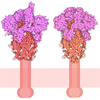
| Title | Structure of the human apo B0AT1-collectrin complex | |||||||||||||||
 Components |
| |||||||||||||||
 Keywords | MEMBRANE PROTEIN / SLC6A19 / collectrin / TMEM27 / apo structure | |||||||||||||||
| Function / homology |  Function and homology information Defective transport of neurotransmitters by SLC6A19 causes Hartnup disorder (HND) / Defective transport of amino acids by SLC6A19 causes Hartnup disorder (HND) / alanine:sodium symporter activity / glycine:sodium symporter activity / calcium-ion regulated exocytosis / neutral amino acid transport / neutral L-amino acid transmembrane transporter activity / SLC-mediated transport of neurotransmitters / Amino acid transport across the plasma membrane / amino acid transmembrane transporter activity ...Defective transport of neurotransmitters by SLC6A19 causes Hartnup disorder (HND) / Defective transport of amino acids by SLC6A19 causes Hartnup disorder (HND) / alanine:sodium symporter activity / glycine:sodium symporter activity / calcium-ion regulated exocytosis / neutral amino acid transport / neutral L-amino acid transmembrane transporter activity / SLC-mediated transport of neurotransmitters / Amino acid transport across the plasma membrane / amino acid transmembrane transporter activity / insulin secretion involved in cellular response to glucose stimulus / Insulin processing / SNARE complex assembly / positive regulation of amino acid transport / amino acid transport / neurotransmitter transport / positive regulation of L-proline import across plasma membrane / transporter activator activity / amino acid import across plasma membrane / viral life cycle / transmembrane transporter complex / response to nutrient / brush border membrane / sodium ion transmembrane transport / apical plasma membrane / protein homodimerization activity / extracellular exosome / plasma membrane / cytoplasm Similarity search - Function | |||||||||||||||
| Biological species |  Homo sapiens (human) | |||||||||||||||
| Method | ELECTRON MICROSCOPY / single particle reconstruction / cryo EM / Resolution: 3.37 Å | |||||||||||||||
 Authors | Pedersen, C.N. / Christensen, M.E. / Heger, T. / Shahsavar, A. / Nissen, P. / Stock, C. | |||||||||||||||
| Funding support |  Denmark, 4items
| |||||||||||||||
 Citation | Journal: J Mol Biol / Year: 2026 Title: Structural Mechanisms of BAT1 Substrate Recognition and Complex Assembly. Authors: Clara N Pedersen / Mads E Christensen / Tomáš Heger / Dheeraj Prakaash / Syma Khalid / Azadeh Shahsavar / Poul Nissen / Charlott Stock /  Abstract: Amino acid transporters are essential for maintaining cellular amino acid homeostasis. The broad-specificity neutral amino acid transporter BAT1 is predominantly expressed in the intestine and ...Amino acid transporters are essential for maintaining cellular amino acid homeostasis. The broad-specificity neutral amino acid transporter BAT1 is predominantly expressed in the intestine and kidney, where it mediates the uptake and reuptake of neutral amino acids with varying affinities. Proper plasma-membrane localization of BAT1 requires dimerization with angiotensin-converting enzyme 2 (ACE2) in the intestine or collectrin in the kidney. Structural information is available for apo and substrate-bound BAT1-ACE2 complexes, but how BAT1 interacts with different accessory proteins and recognizes smaller substrates remains enigmatic. Here, we compare cryo-EM structures of BAT1 in complex with ACE2 or collectrin in substrate-free states, and bound to alanine, leucine or tryptophan. The complexes display only minor differences in accessory-protein binding through a conserved interaction surface. Substrate recognition follows a canonical architecture observed for other SLC6 family transporters, with subtle adaptations that potentially account for the different affinities of the three amino acids. Notably, we resolve several cholesterol molecules surrounding the transporter, highlighting a potential regulatory role of the membrane environment that we address by molecular dynamics simulations. | |||||||||||||||
| History |
|
- Structure visualization
| Structure viewer | Molecule: MolmilJmol/JSmol |
|---|
- Downloads & links
-Download
| PDBx/mmCIF format | 34oa.cif.gz | 612.5 KB | Display | PDBx/mmCIF format |
|---|---|---|---|---|
| PDB format | pdb34oa.ent.gz | 523.5 KB | Display | PDB format |
| PDBx/mmJSON format | 34oa.json.gz | Tree view | PDBx/mmJSON format | |
| Others |  Other downloads |
-Validation report
| Arichive directory | https://data.pdbj.org/pub/pdb/validation_reports/4o/34oaftp://data.pdbj.org/pub/pdb/validation_reports/4o/34oa | HTTPS FTP |
|---|
-Related structure data
| Related structure data |  100048MC  34ocC  34odC  34oeC  34ohC C: citing same article ( M: map data used to model this data |
|---|---|
| Similar structure data |


-Links
 PDBj

- Assembly
| Deposited unit | 
|
|---|---|
| 1 |
|
-Components
-Protein , 2 types, 4 molecules ABCD
| #1: Protein | Mass: 72150.406 Da / Num. of mol.: 2 Source method: isolated from a genetically manipulated source Source: (gene. exp.) Homo sapiens (human) / Gene: SLC6A19, B0AT1 / Production host: Homo sapiens (human) / References: UniProt: Q695T7#2: Protein | Mass: 26292.523 Da / Num. of mol.: 2 Source method: isolated from a genetically manipulated source Source: (gene. exp.) Homo sapiens (human) / Gene: CLTRN, TMEM27, UNQ679/PRO1312 / Production host: Homo sapiens (human) / References: UniProt: Q9HBJ8 |
|---|
-Sugars , 3 types, 16 molecules 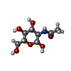

| #3: Polysaccharide | Source method: isolated from a genetically manipulated source #4: Sugar | ChemComp-NAG /  Type: D-saccharide, beta linking / Mass: 221.208 Da / Num. of mol.: 12 / Source method: obtained synthetically / Formula: C8H15NO6 #6: Sugar |  Type: D-saccharide, alpha linking / Mass: 221.208 Da / Num. of mol.: 2 / Source method: obtained synthetically / Formula: C8H15NO6 |
|---|
-Non-polymers , 1 types, 16 molecules 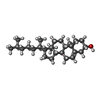
| #5: Chemical | ChemComp-CLR /  Mass: 386.654 Da / Num. of mol.: 16 / Source method: obtained synthetically / Formula: C27H46O |
|---|
-Details
| Has ligand of interest | N |
|---|---|
| Has protein modification | Y |
-Experimental details
-Experiment
| Experiment | Method: ELECTRON MICROSCOPY |
|---|---|
| EM experiment | Aggregation state: PARTICLE / 3D reconstruction method: single particle reconstruction |
- Sample preparation
| Component | Name: dimer of heterodimer between B0AT1-collectrin / Type: COMPLEX / Entity ID: #1-#2 / Source: RECOMBINANT |
|---|---|
| Molecular weight | Value: 0.193 MDa / Experimental value: NO |
| Source (natural) | Organism: Homo sapiens (human) |
| Source (recombinant) | Organism: Homo sapiens (human) |
| Buffer solution | pH: 8 |
| Specimen | Conc.: 1.2 mg/ml / Embedding applied: NO / Shadowing applied: NO / Staining applied: NO / Vitrification applied: YES |
| Specimen support | Grid material: GOLD / Grid mesh size: 300 divisions/in. / Grid type: UltrAuFoil R0./1 |
| Vitrification | Instrument: FEI VITROBOT MARK IV / Cryogen name: ETHANE / Humidity: 100 % / Chamber temperature: 277 K |
- Electron microscopy imaging
| Experimental equipment |  Model: Titan Krios / Image courtesy: FEI Company |
|---|---|
| Microscopy | Model: TFS KRIOS |
| Electron gun | Electron source:  FIELD EMISSION GUN / Accelerating voltage: 300 kV / Illumination mode: FLOOD BEAM |
| Electron lens | Mode: BRIGHT FIELD / Nominal magnification: 130000 X / Nominal defocus max: 2000 nm / Nominal defocus min: 800 nm / Cs: 2.7 mm |
| Specimen holder | Specimen holder model: FEI TITAN KRIOS AUTOGRID HOLDER |
| Image recording | Electron dose: 59.7 e/Å2 / Film or detector model: GATAN K3 (6k x 4k) / Num. of real images: 6374 |
- Processing
| EM software |
| |||||||||||||||||||||||||||||||||
|---|---|---|---|---|---|---|---|---|---|---|---|---|---|---|---|---|---|---|---|---|---|---|---|---|---|---|---|---|---|---|---|---|---|---|
| CTF correction | Type: PHASE FLIPPING AND AMPLITUDE CORRECTION | |||||||||||||||||||||||||||||||||
| Particle selection | Num. of particles selected: 199212 | |||||||||||||||||||||||||||||||||
| 3D reconstruction | Resolution: 3.37 Å / Resolution method: FSC 0.143 CUT-OFF / Num. of particles: 88151 / Symmetry type: POINT | |||||||||||||||||||||||||||||||||
| Atomic model building | Protocol: FLEXIBLE FIT Details: Initial rigid body fitting and then flexible fitting using Namdinator.au.dk | |||||||||||||||||||||||||||||||||
| Atomic model building |
|