Movie
 Controller


[English] 日本語
 Yorodumi
- EMDB-100018: Focussed map of the transmembrane region of B0AT1-ACE2 complex bo... -
+ Open data

- Basic information
| Entry |  | |||||||||||||||
|---|---|---|---|---|---|---|---|---|---|---|---|---|---|---|---|---|
| Title | Focussed map of the transmembrane region of B0AT1-ACE2 complex bound to L-Tryptophan | |||||||||||||||
 Map data | ||||||||||||||||
 Sample |
| |||||||||||||||
 Keywords | B0AT1-ACE2 complex / substrate binding / cholesterol binding / occluded / sodium binding site / MEMBRANE PROTEIN | |||||||||||||||
| Biological species |  Homo sapiens (human) | |||||||||||||||
| Method | single particle reconstruction / cryo EM / Resolution: 3.12 Å | |||||||||||||||
 Authors | Pedersen CN / Christensen ME / Heger T / Shahsavar A / Nissen P / Stock C | |||||||||||||||
| Funding support |  Denmark, 4 items
| |||||||||||||||
 Citation | Journal: J Mol Biol / Year: 2026 Title: Structural Mechanisms of BAT1 Substrate Recognition and Complex Assembly. Authors: Clara N Pedersen / Mads E Christensen / Tomáš Heger / Dheeraj Prakaash / Syma Khalid / Azadeh Shahsavar / Poul Nissen / Charlott Stock /  Abstract: Amino acid transporters are essential for maintaining cellular amino acid homeostasis. The broad-specificity neutral amino acid transporter BAT1 is predominantly expressed in the intestine and ...Amino acid transporters are essential for maintaining cellular amino acid homeostasis. The broad-specificity neutral amino acid transporter BAT1 is predominantly expressed in the intestine and kidney, where it mediates the uptake and reuptake of neutral amino acids with varying affinities. Proper plasma-membrane localization of BAT1 requires dimerization with angiotensin-converting enzyme 2 (ACE2) in the intestine or collectrin in the kidney. Structural information is available for apo and substrate-bound BAT1-ACE2 complexes, but how BAT1 interacts with different accessory proteins and recognizes smaller substrates remains enigmatic. Here, we compare cryo-EM structures of BAT1 in complex with ACE2 or collectrin in substrate-free states, and bound to alanine, leucine or tryptophan. The complexes display only minor differences in accessory-protein binding through a conserved interaction surface. Substrate recognition follows a canonical architecture observed for other SLC6 family transporters, with subtle adaptations that potentially account for the different affinities of the three amino acids. Notably, we resolve several cholesterol molecules surrounding the transporter, highlighting a potential regulatory role of the membrane environment that we address by molecular dynamics simulations. | |||||||||||||||
| History |
|
- Structure visualization
| Supplemental images |
|---|

- Downloads & links
-EMDB archive
| Map data | emd_100018.map.gz | 60.4 MB |  EMDB map data format | |
|---|---|---|---|---|
| Header (meta data) | emd-100018-v30.xmlemd-100018.xml | 20.4 KB 20.4 KB | Display Display | EMDB header |
| FSC (resolution estimation) | emd_100018_fsc.xml | 10.6 KB | Display | FSC data file |
| Images |  emd_100018.png | 38.1 KB | ||
| Masks | emd_100018_msk_1.map | 125 MB | Mask map | |
| Filedesc metadata | emd-100018.cif.gz | 6.5 KB | ||
| Others | emd_100018_half_map_1.map.gzemd_100018_half_map_2.map.gz | 114.9 MB 114.9 MB | ||
| Archive directory |  https://data.pdbj.org/pub/emdb/structures/EMD-100018ftp://data.pdbj.org/pub/emdb/structures/EMD-100018 | HTTPS FTP |
-Related structure data


-Links
| EMDB pages | EMDB (EBI/PDBe) / EMDataResource |
|---|
-Map
| File | Download / File: emd_100018.map.gz / Format: CCP4 / Size: 125 MB / Type: IMAGE STORED AS FLOATING POINT NUMBER (4 BYTES) | ||||||||||||||||||||||||||||||||||||
|---|---|---|---|---|---|---|---|---|---|---|---|---|---|---|---|---|---|---|---|---|---|---|---|---|---|---|---|---|---|---|---|---|---|---|---|---|---|
| Projections & slices | Image control
Images are generated by Spider. | ||||||||||||||||||||||||||||||||||||
| Voxel size | X=Y=Z: 1.0352 Å | ||||||||||||||||||||||||||||||||||||
| Density |
| ||||||||||||||||||||||||||||||||||||
| Symmetry | Space group: 1 | ||||||||||||||||||||||||||||||||||||
| Details | EMDB XML:
|
 Z (Sec.)
 Y (Row.)
 X (Col.)


-Supplemental data
-Mask #1
| File | emd_100018_msk_1.map | ||||||||||||
|---|---|---|---|---|---|---|---|---|---|---|---|---|---|
| Projections & Slices |
| ||||||||||||
| Density Histograms |


-Half map: #1
| File | emd_100018_half_map_1.map | ||||||||||||
|---|---|---|---|---|---|---|---|---|---|---|---|---|---|
| Projections & Slices |
| ||||||||||||
| Density Histograms |


-Half map: #2
| File | emd_100018_half_map_2.map | ||||||||||||
|---|---|---|---|---|---|---|---|---|---|---|---|---|---|
| Projections & Slices |
| ||||||||||||
| Density Histograms |


- Sample components
-Entire : dimer of heterodimer between B0AT1-ACE2
| Entire | Name: dimer of heterodimer between B0AT1-ACE2 |
|---|---|
| Components |
|
-Supramolecule #1: dimer of heterodimer between B0AT1-ACE2
| Supramolecule | Name: dimer of heterodimer between B0AT1-ACE2 / type: complex / ID: 1 / Parent: 0 / Macromolecule list: all |
|---|---|
| Source (natural) | Organism: Homo sapiens (human) |
| Molecular weight | Theoretical: 327 KDa |
-Macromolecule #1: B0AT1, SLC6A19
| Macromolecule | Name: B0AT1, SLC6A19 / type: protein_or_peptide / ID: 1 / Number of copies: 2 / Enantiomer: LEVO |
|---|---|
| Source (natural) | Organism: Homo sapiens (human) |
| Molecular weight | Theoretical: 68.11575 KDa |
| Recombinant expression | Organism: Homo sapiens (human) |
| Sequence | String: MVRLVLPNPG LDARIPSLAE LETIEQEEAS SRPKWDNKAQ YMLTCLGFCV GLGNVWRFPY LCQSHGGGA FMIPFLILLV LEGIPLLYLE FAIGQRLRRG SLGVWSSIHP ALKGLGLASM L TSFMVGLY YNTIISWIMW YLFNSFQEPL PWSDCPLNEN QTGYVDECAR ...String: MVRLVLPNPG LDARIPSLAE LETIEQEEAS SRPKWDNKAQ YMLTCLGFCV GLGNVWRFPY LCQSHGGGA FMIPFLILLV LEGIPLLYLE FAIGQRLRRG SLGVWSSIHP ALKGLGLASM L TSFMVGLY YNTIISWIMW YLFNSFQEPL PWSDCPLNEN QTGYVDECAR SSPVDYFWYR ET LNISTSI SDSGSIQWWM LLCLACAWSV LYMCTIRGIE TTGKAVYITS TLPYVVLTIF LIR GLTLKG ATNGIVFLFT PNVTELAQPD TWLDAGAQVF FSFSLAFGGL ISFSSYNSVH NNCE KDSVI VSIINGFTSV YVAIVVYSVI GFRATQRYDD CFSTNILTLI NGFDLPEGNV TQENF VDMQ QRCNASDPAA YAQLVFQTCD INAFLSEAVE GTGLAFIVFT EAITKMPLSP LWSVLF FIM LFCLGLSSMF GNMEGVVVPL QDLRVIPPKW PKEVLTGLIC LGTFLIGFIF TLNSGQY WL SLLDSYAGSI PLLIIAFCEM FSVVYVYGVD RFNKDIEFMI GHKPNIFWQV TWRVVSPL L MLIIFLFFFV VEVSQELTYS IWDPGYEEFP KSQKISYPNW VYVVVVIVAG VPSLTIPGY AIYKLIRNHC QKPGDHQGLV STLSTASMNG DLKYGGRLEV LFQ |
-Macromolecule #2: ACE2
| Macromolecule | Name: ACE2 / type: protein_or_peptide / ID: 2 / Number of copies: 2 / Enantiomer: LEVO |
|---|---|
| Source (natural) | Organism: Homo sapiens (human) |
| Molecular weight | Theoretical: 87.078734 KDa |
| Recombinant expression | Organism: Homo sapiens (human) |
| Sequence | String: MSSSSWLLLS LVAVTAAQST IEEQAKTFLD KFNHEAEDLF YQSSLASWNY NTNITEENVQ NMNNAGDKW SAFLKEQSTL AQMYPLQEIQ NLTVKLQLQA LQQNGSSVLS EDKSKRLNTI L NTMSTIYS TGKVCNPDNP QECLLLEPGL NEIMANSLDY NERLWAWESW ...String: MSSSSWLLLS LVAVTAAQST IEEQAKTFLD KFNHEAEDLF YQSSLASWNY NTNITEENVQ NMNNAGDKW SAFLKEQSTL AQMYPLQEIQ NLTVKLQLQA LQQNGSSVLS EDKSKRLNTI L NTMSTIYS TGKVCNPDNP QECLLLEPGL NEIMANSLDY NERLWAWESW RSEVGKQLRP LY EEYVVLK NEMARANHYE DYGDYWRGDY EVNGVDGYDY SRGQLIEDVE HTFEEIKPLY EHL HAYVRA KLMNAYPSYI SPIGCLPAHL LGDMWGRFWT NLYSLTVPFG QKPNIDVTDA MVDQ AWDAQ RIFKEAEKFF VSVGLPNMTQ GFWENSMLTD PGNVQKAVCH PTAWDLGKGD FRILM CTKV TMDDFLTAHH EMGHIQYDMA YAAQPFLLRN GANEGFHEAV GEIMSLSAAT PKHLKS IGL LSPDFQEDNE TEINFLLKQA LTIVGTLPFT YMLEKWRWMV FKGEIPKDQW MKKWWEM KR EIVGVVEPVP HDETYCDPAS LFHVSNDYSF IRYYTRTLYQ FQFQEALCQA AKHEGPLH K CDISNSTEAG QKLFNMLRLG KSEPWTLALE NVVGAKNMNV RPLLNYFEPL FTWLKDQNK NSFVGWSTDW SPYADQSIKV RISLKSALGD RAYEWNDNEM YLFRSSVAYA MRQYFLKVKN QMILFGEED VRVANLKPRI SFNFFVTAPK NVSDIIPRTE VEKAIRMSRS RINDAFRLND N SLEFLGIQ PTLGPPNQPP VSIWLIVFGV VMGVIVVGIV ILIFTGIRDR KKKNKARSGE NP YASIDIS KGENNPGFQN TDDVQTSFSR LEVLFQ |
-Experimental details
-Structure determination
| Method | cryo EM |
|---|---|
 Processing | single particle reconstruction |
| Aggregation state | particle |
-Sample preparation
| Concentration | 1.2 mg/mL |
|---|---|
| Buffer | pH: 8 |
| Grid | Model: UltrAuFoil R0./1 / Material: GOLD / Mesh: 300 / Pretreatment - Type: GLOW DISCHARGE / Pretreatment - Time: 90 sec. |
| Vitrification | Cryogen name: ETHANE / Chamber humidity: 100 % / Chamber temperature: 277 K / Instrument: FEI VITROBOT MARK IV |
- Electron microscopy
| Microscope | TFS KRIOS |
|---|---|
| Software | Name: EPU |
| Image recording | Film or detector model: GATAN K3 (6k x 4k) / Number real images: 6215 / Average electron dose: 59.9 e/Å2 |
| Electron beam | Acceleration voltage: 300 kV / Electron source:  FIELD EMISSION GUN |
| Electron optics | Illumination mode: FLOOD BEAM / Imaging mode: BRIGHT FIELD / Cs: 2.7 mm / Nominal defocus max: 2.0 µm / Nominal defocus min: 0.8 µm / Nominal magnification: 130000 |
| Sample stage | Specimen holder model: FEI TITAN KRIOS AUTOGRID HOLDER / Cooling holder cryogen: NITROGEN |
| Experimental equipment | 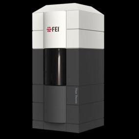 Model: Titan Krios / Image courtesy: FEI Company |