Movie
 Controller


[English] 日本語
 Yorodumi
- EMDB-100015: Focussed map of the extracellular region of B0AT1-ACE2 apo complex -
+ Open data

- Basic information
| Entry |  | |||||||||||||||
|---|---|---|---|---|---|---|---|---|---|---|---|---|---|---|---|---|
| Title | Focussed map of the extracellular region of B0AT1-ACE2 apo complex | |||||||||||||||
 Map data | ||||||||||||||||
 Sample |
| |||||||||||||||
 Keywords | SLC6A19 / ACE2 / apo broad neutral amino acid transporter / extracellular region / MEMBRANE PROTEIN | |||||||||||||||
| Biological species |  Homo Sapiens (human) / Homo sapiens (human) | |||||||||||||||
| Method | single particle reconstruction / cryo EM / Resolution: 2.98 Å | |||||||||||||||
 Authors | Pedersen CN / Christensen ME / Heger T / Shahsavar A / Nissen P / Stock C | |||||||||||||||
| Funding support |  Denmark, 4 items
| |||||||||||||||
 Citation | Journal: J Mol Biol / Year: 2026 Title: Structural Mechanisms of BAT1 Substrate Recognition and Complex Assembly. Authors: Clara N Pedersen / Mads E Christensen / Tomáš Heger / Dheeraj Prakaash / Syma Khalid / Azadeh Shahsavar / Poul Nissen / Charlott Stock /  Abstract: Amino acid transporters are essential for maintaining cellular amino acid homeostasis. The broad-specificity neutral amino acid transporter BAT1 is predominantly expressed in the intestine and ...Amino acid transporters are essential for maintaining cellular amino acid homeostasis. The broad-specificity neutral amino acid transporter BAT1 is predominantly expressed in the intestine and kidney, where it mediates the uptake and reuptake of neutral amino acids with varying affinities. Proper plasma-membrane localization of BAT1 requires dimerization with angiotensin-converting enzyme 2 (ACE2) in the intestine or collectrin in the kidney. Structural information is available for apo and substrate-bound BAT1-ACE2 complexes, but how BAT1 interacts with different accessory proteins and recognizes smaller substrates remains enigmatic. Here, we compare cryo-EM structures of BAT1 in complex with ACE2 or collectrin in substrate-free states, and bound to alanine, leucine or tryptophan. The complexes display only minor differences in accessory-protein binding through a conserved interaction surface. Substrate recognition follows a canonical architecture observed for other SLC6 family transporters, with subtle adaptations that potentially account for the different affinities of the three amino acids. Notably, we resolve several cholesterol molecules surrounding the transporter, highlighting a potential regulatory role of the membrane environment that we address by molecular dynamics simulations. | |||||||||||||||
| History |
|
- Structure visualization
| Supplemental images |
|---|

- Downloads & links
-EMDB archive
| Map data | emd_100015.map.gz | 52 MB |  EMDB map data format | |
|---|---|---|---|---|
| Header (meta data) | emd-100015-v30.xmlemd-100015.xml | 20.1 KB 20.1 KB | Display Display | EMDB header |
| FSC (resolution estimation) | emd_100015_fsc.xml | 9.8 KB | Display | FSC data file |
| Images |  emd_100015.png | 46.4 KB | ||
| Masks | emd_100015_msk_1.map | 103 MB | Mask map | |
| Filedesc metadata | emd-100015.cif.gz | 6.4 KB | ||
| Others | emd_100015_half_map_1.map.gzemd_100015_half_map_2.map.gz | 95.4 MB 95.4 MB | ||
| Archive directory |  https://data.pdbj.org/pub/emdb/structures/EMD-100015ftp://data.pdbj.org/pub/emdb/structures/EMD-100015 | HTTPS FTP |
-Related structure data


-Links
| EMDB pages | EMDB (EBI/PDBe) / EMDataResource |
|---|
-Map
| File | Download / File: emd_100015.map.gz / Format: CCP4 / Size: 103 MB / Type: IMAGE STORED AS FLOATING POINT NUMBER (4 BYTES) | ||||||||||||||||||||||||||||||||||||
|---|---|---|---|---|---|---|---|---|---|---|---|---|---|---|---|---|---|---|---|---|---|---|---|---|---|---|---|---|---|---|---|---|---|---|---|---|---|
| Projections & slices | Image control
Images are generated by Spider. | ||||||||||||||||||||||||||||||||||||
| Voxel size | X=Y=Z: 1.294 Å | ||||||||||||||||||||||||||||||||||||
| Density |
| ||||||||||||||||||||||||||||||||||||
| Symmetry | Space group: 1 | ||||||||||||||||||||||||||||||||||||
| Details | EMDB XML:
|
 Z (Sec.)
 Y (Row.)
 X (Col.)


-Supplemental data
-Mask #1
| File | emd_100015_msk_1.map | ||||||||||||
|---|---|---|---|---|---|---|---|---|---|---|---|---|---|
| Projections & Slices |
| ||||||||||||
| Density Histograms |


-Half map: #1
| File | emd_100015_half_map_1.map | ||||||||||||
|---|---|---|---|---|---|---|---|---|---|---|---|---|---|
| Projections & Slices |
| ||||||||||||
| Density Histograms |


-Half map: #2
| File | emd_100015_half_map_2.map | ||||||||||||
|---|---|---|---|---|---|---|---|---|---|---|---|---|---|
| Projections & Slices |
| ||||||||||||
| Density Histograms |


- Sample components
-Entire : dimer of heterodimer between B0AT1-ACE2
| Entire | Name: dimer of heterodimer between B0AT1-ACE2 |
|---|---|
| Components |
|
-Supramolecule #1: dimer of heterodimer between B0AT1-ACE2
| Supramolecule | Name: dimer of heterodimer between B0AT1-ACE2 / type: complex / ID: 1 / Parent: 0 / Macromolecule list: all |
|---|---|
| Source (natural) | Organism: Homo Sapiens (human) |
| Molecular weight | Theoretical: 327 KDa |
-Macromolecule #1: B0AT1, SLC6A19
| Macromolecule | Name: B0AT1, SLC6A19 / type: protein_or_peptide / ID: 1 / Number of copies: 2 / Enantiomer: LEVO |
|---|---|
| Source (natural) | Organism: Homo sapiens (human) |
| Molecular weight | Theoretical: 72.150406 KDa |
| Recombinant expression | Organism: Homo sapiens (human) |
| Sequence | String: MVRLVLPNPG LDARIPSLAE LETIEQEEAS SRPKWDNKAQ YMLTCLGFCV GLGNVWRFPY LCQSHGGGAF MIPFLILLVL EGIPLLYLE FAIGQRLRRG SLGVWSSIHP ALKGLGLASM LTSFMVGLYY NTIISWIMWY LFNSFQEPLP WSDCPLNENQ T GYVDECAR ...String: MVRLVLPNPG LDARIPSLAE LETIEQEEAS SRPKWDNKAQ YMLTCLGFCV GLGNVWRFPY LCQSHGGGAF MIPFLILLVL EGIPLLYLE FAIGQRLRRG SLGVWSSIHP ALKGLGLASM LTSFMVGLYY NTIISWIMWY LFNSFQEPLP WSDCPLNENQ T GYVDECAR SSPVDYFWYR ETLNISTSIS DSGSIQWWML LCLACAWSVL YMCTIRGIET TGKAVYITST LPYVVLTIFL IR GLTLKGA TNGIVFLFTP NVTELAQPDT WLDAGAQVFF SFSLAFGGLI SFSSYNSVHN NCEKDSVIVS IINGFTSVYV AIV VYSVIG FRATQRYDDC FSTNILTLIN GFDLPEGNVT QENFVDMQQR CNASDPAAYA QLVFQTCDIN AFLSEAVEGT GLAF IVFTE AITKMPLSPL WSVLFFIMLF CLGLSSMFGN MEGVVVPLQD LRVIPPKWPK EVLTGLICLG TFLIGFIFTL NSGQY WLSL LDSYAGSIPL LIIAFCEMFS VVYVYGVDRF NKDIEFMIGH KPNIFWQVTW RVVSPLLMLI IFLFFFVVEV SQELTY SIW DPGYEEFPKS QKISYPNWVY VVVVIVAGVP SLTIPGYAIY KLIRNHCQKP GDHQGLVSTL STASMNGDLK YGGRLEV LF Q |
-Macromolecule #2: ACE2
| Macromolecule | Name: ACE2 / type: protein_or_peptide / ID: 2 / Number of copies: 2 / Enantiomer: LEVO |
|---|---|
| Source (natural) | Organism: Homo sapiens (human) |
| Molecular weight | Theoretical: 93.558844 KDa |
| Recombinant expression | Organism: Homo sapiens (human) |
| Sequence | String: MSSSSWLLLS LVAVTAAQST IEEQAKTFLD KFNHEAEDLF YQSSLASWNY NTNITEENVQ NMNNAGDKWS AFLKEQSTLA QMYPLQEIQ NLTVKLQLQA LQQNGSSVLS EDKSKRLNTI LNTMSTIYST GKVCNPDNPQ ECLLLEPGLN EIMANSLDYN E RLWAWESW ...String: MSSSSWLLLS LVAVTAAQST IEEQAKTFLD KFNHEAEDLF YQSSLASWNY NTNITEENVQ NMNNAGDKWS AFLKEQSTLA QMYPLQEIQ NLTVKLQLQA LQQNGSSVLS EDKSKRLNTI LNTMSTIYST GKVCNPDNPQ ECLLLEPGLN EIMANSLDYN E RLWAWESW RSEVGKQLRP LYEEYVVLKN EMARANHYED YGDYWRGDYE VNGVDGYDYS RGQLIEDVEH TFEEIKPLYE HL HAYVRAK LMNAYPSYIS PIGCLPAHLL GDMWGRFWTN LYSLTVPFGQ KPNIDVTDAM VDQAWDAQRI FKEAEKFFVS VGL PNMTQG FWENSMLTDP GNVQKAVCHP TAWDLGKGDF RILMCTKVTM DDFLTAHHEM GHIQYDMAYA AQPFLLRNGA NEGF HEAVG EIMSLSAATP KHLKSIGLLS PDFQEDNETE INFLLKQALT IVGTLPFTYM LEKWRWMVFK GEIPKDQWMK KWWEM KREI VGVVEPVPHD ETYCDPASLF HVSNDYSFIR YYTRTLYQFQ FQEALCQAAK HEGPLHKCDI SNSTEAGQKL FNMLRL GKS EPWTLALENV VGAKNMNVRP LLNYFEPLFT WLKDQNKNSF VGWSTDWSPY ADQSIKVRIS LKSALGDRAY EWNDNEM YL FRSSVAYAMR QYFLKVKNQM ILFGEEDVRV ANLKPRISFN FFVTAPKNVS DIIPRTEVEK AIRMSRSRIN DAFRLNDN S LEFLGIQPTL GPPNQPPVSI WLIVFGVVMG VIVVGIVILI FTGIRDRKKK NKARSGENPY ASIDISKGEN NPGFQNTDD VQTSFSRLEV LFQ |
-Experimental details
-Structure determination
| Method | cryo EM |
|---|---|
 Processing | single particle reconstruction |
| Aggregation state | particle |
-Sample preparation
| Concentration | 1.2 mg/mL |
|---|---|
| Buffer | pH: 8 |
| Grid | Model: UltrAuFoil R0./1 / Material: GOLD / Mesh: 300 / Pretreatment - Type: GLOW DISCHARGE / Pretreatment - Time: 90 sec. |
| Vitrification | Cryogen name: ETHANE / Chamber humidity: 100 % / Chamber temperature: 277 K / Instrument: FEI VITROBOT MARK IV |
- Electron microscopy
| Microscope | TFS KRIOS |
|---|---|
| Image recording | Film or detector model: GATAN K3 (6k x 4k) / Number real images: 10763 / Average electron dose: 59.5 e/Å2 |
| Electron beam | Acceleration voltage: 300 kV / Electron source:  FIELD EMISSION GUN |
| Electron optics | Illumination mode: FLOOD BEAM / Imaging mode: BRIGHT FIELD / Cs: 2.7 mm / Nominal defocus max: 2.0 µm / Nominal defocus min: 0.8 µm / Nominal magnification: 130000 |
| Sample stage | Specimen holder model: FEI TITAN KRIOS AUTOGRID HOLDER / Cooling holder cryogen: NITROGEN |
| Experimental equipment | 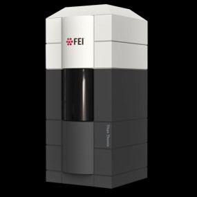 Model: Titan Krios / Image courtesy: FEI Company |