 Movie
Movie Controller


+ Open data

- Basic information
| Entry | Database: PDB / ID: 31ak | ||||||||||||
|---|---|---|---|---|---|---|---|---|---|---|---|---|---|
| Title | Structure of native human leukocyte myeloperoxidase | ||||||||||||
 Components Components |
| ||||||||||||
 Keywords Keywords | OXIDOREDUCTASE / Innate immunity / neutrophils / phagocytosis / NETosis | ||||||||||||
| Function / homology |  Function and homology information Function and homology informationEvents associated with phagocytolytic activity of PMN cells / neutrophil-mediated killing of symbiont cell / myeloperoxidase / positive regulation of cholesterol import / neutrophil extracellular trap / phagocytic vesicle lumen / neutrophil-mediated killing of bacterium / cytolytic granule / response to gold nanoparticle / thiocyanate peroxidase activity ...Events associated with phagocytolytic activity of PMN cells / neutrophil-mediated killing of symbiont cell / myeloperoxidase / positive regulation of cholesterol import / neutrophil extracellular trap / phagocytic vesicle lumen / neutrophil-mediated killing of bacterium / cytolytic granule / response to gold nanoparticle / thiocyanate peroxidase activity / neutrophil extracellular trap formation / neutrophil-mediated killing of fungus / protein-containing complex destabilizing activity / low-density lipoprotein particle remodeling / nucleosome disassembly / azurophil granule / response to food / response to mechanical stimulus / phagocytic vesicle / nucleosome binding / secretory granule / hydrogen peroxide catabolic process / peroxidase activity / defense response / azurophil granule lumen / heparin binding / response to lipopolysaccharide / response to oxidative stress / defense response to bacterium / lysosome / chromosome / chromatin binding / heme binding / negative regulation of apoptotic process / Neutrophil degranulation / extracellular exosome / extracellular region / nucleus Similarity search - Function | ||||||||||||
| Biological species |  Homo sapiens (human) Homo sapiens (human) | ||||||||||||
| Method | ELECTRON MICROSCOPY / single particle reconstruction / cryo EM / Resolution: 2.18 Å | ||||||||||||
 Authors Authors | Leitgeb, U. / Pfanzagl, V. / Guo, Y. / Emde, T. / Borek, D. | ||||||||||||
| Funding support |  Austria, 3items Austria, 3items
| ||||||||||||
 Citation Citation | Journal: Int J Biol Macromol / Year: 2026 Title: Interface N-glycans drive myeloperoxidase dimerization in vitro. Authors: Urban Leitgeb / Yirui Guo / Tabitha Emde / Valentina Ruocco / Theodor Simak / Ela Zdenkovic / Paul G Furtmüller / Dominika Borek / William M Nauseef / Chris Oostenbrink / Vera Pfanzagl /   Abstract: Myeloperoxidase (MPO) is a highly glycosylated heme oxidoreductase that, together with lactoperoxidase (LPO) and eosinophil peroxidase (EPO), contributes to host defence through the generation of ...Myeloperoxidase (MPO) is a highly glycosylated heme oxidoreductase that, together with lactoperoxidase (LPO) and eosinophil peroxidase (EPO), contributes to host defence through the generation of (pseudo)hypohalous acids. Among the human heme peroxidases, mature MPO forms a unique covalently linked homodimer. Dimerization enhances stability and is essential for some biological functions, including chromatin disruption during neutrophil extracellular trap formation. Thus, we hypothesized that MPO dimerization must reflect a tightly regulated step in MPO biosynthesis and is driven by specific structural elements. Cross-species sequence alignment indicated that N-glycosylation motifs were evolutionarily more conserved in MPO than in monomeric EPO and LPO. We investigated the role of N-glycans in MPO dimerization using in vitro monomerized glycosylated (mMPO) and deglycosylated (mMPOdg) monomeric variants of native human MPO. Small-angle X-ray scattering, isothermal titration calorimetry and crystal structures showed that glycosylated MPO monomers had a weak but biologically relevant affinity in the millimolar range, which was lost upon deglycosylation. Furthermore, we present the first cryogenic transmission electron microscopy structure of human MPO with at least one core N-acetylglucosamine residue resolved at each N-glycosylation site. Structural analysis suggests that an extensive hydrogen bonding network of interface N-glycans at position 483 and residues of the other monomer drives MPO homodimerization. Dimerization enhances stability without affecting enzymatic activity. Together, we show that N-glycans are essential to MPO dimerization, provide important insights into the impact of interface N-glycans on biophysical and biochemical properties of MPO and discuss the implications for the biological roles of MPO in health and disease. | ||||||||||||
| History |
|
- Structure visualization
| Structure viewer | Molecule: MolmilJmol/JSmol |
|---|
- Downloads & links
-Download
| PDBx/mmCIF format | 31ak.cif.gz | 227.9 KB | Display | PDBx/mmCIF format |
|---|---|---|---|---|
| PDB format | pdb31ak.ent.gz | 177.5 KB | Display | PDB format |
| PDBx/mmJSON format | 31ak.json.gz | Tree view | PDBx/mmJSON format | |
| Others |  Other downloads Other downloads |
-Validation report
| Arichive directory | https://data.pdbj.org/pub/pdb/validation_reports/1a/31akftp://data.pdbj.org/pub/pdb/validation_reports/1a/31ak | HTTPS FTP |
|---|
-Related structure data
| Related structure data |  58198MC  29toC  29wjC M: map data used to model this data C: citing same article ( |
|---|---|
| Similar structure data |
-Links
 PDBj
PDBj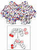
- Assembly
| Deposited unit | 
|
|---|---|
| 1 |
|
-Components
-Protein , 2 types, 4 molecules AEBF
| #1: Protein | Mass: 12894.465 Da / Num. of mol.: 2 / Fragment: UNP RESIDUES 167-271 / Source method: isolated from a natural source / Source: (natural) Homo sapiens (human) / References: UniProt: P05164#2: Protein | Mass: 53305.266 Da / Num. of mol.: 2 / Source method: isolated from a natural source / Source: (natural) Homo sapiens (human) / References: UniProt: P05164, myeloperoxidase |
|---|
-Sugars , 5 types, 10 molecules 
| #3: Polysaccharide | Source method: isolated from a genetically manipulated source #4: Polysaccharide | Source method: isolated from a genetically manipulated source #5: Polysaccharide | alpha-D-mannopyranose-(1-3)-[alpha-D-mannopyranose-(1-6)]beta-D-mannopyranose-(1-4)-2-acetamido-2- ...alpha-D-mannopyranose-(1-3)-[alpha-D-mannopyranose-(1-6)]beta-D-mannopyranose-(1-4)-2-acetamido-2-deoxy-beta-D-glucopyranose-(1-4)-[alpha-L-fucopyranose-(1-6)]2-acetamido-2-deoxy-beta-D-glucopyranose | Source method: isolated from a genetically manipulated source #6: Polysaccharide | alpha-D-mannopyranose-(1-6)-alpha-D-mannopyranose-(1-6)-[alpha-D-mannopyranose-(1-3)]beta-D- ...alpha-D-mannopyranose-(1-6)-alpha-D-mannopyranose-(1-6)-[alpha-D-mannopyranose-(1-3)]beta-D-mannopyranose-(1-4)-2-acetamido-2-deoxy-beta-D-glucopyranose-(1-4)-[alpha-L-fucopyranose-(1-6)]2-acetamido-2-deoxy-beta-D-glucopyranose | Type: oligosaccharide / Mass: 1219.105 Da / Num. of mol.: 1 Source method: isolated from a genetically manipulated source #10: Sugar | ChemComp-NAG /  Type: D-saccharide, beta linking / Mass: 221.208 Da / Num. of mol.: 4 / Source method: obtained synthetically / Formula: C8H15NO6 Type: D-saccharide, beta linking / Mass: 221.208 Da / Num. of mol.: 4 / Source method: obtained synthetically / Formula: C8H15NO6 |
|---|
-Non-polymers , 3 types, 8 molecules 

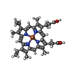
| #7: Chemical | ChemComp-CL /  Mass: 35.453 Da / Num. of mol.: 4 / Source method: obtained synthetically / Formula: Cl Mass: 35.453 Da / Num. of mol.: 4 / Source method: obtained synthetically / Formula: Cl#8: Chemical |  Mass: 40.078 Da / Num. of mol.: 2 / Source method: obtained synthetically / Formula: Ca Mass: 40.078 Da / Num. of mol.: 2 / Source method: obtained synthetically / Formula: Ca#9: Chemical | 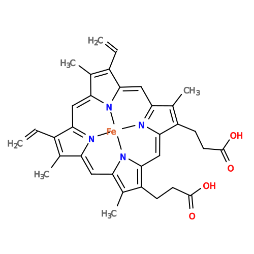 Mass: 616.487 Da / Num. of mol.: 2 / Source method: obtained synthetically / Formula: C34H32FeN4O4 / Feature type: SUBJECT OF INVESTIGATION Mass: 616.487 Da / Num. of mol.: 2 / Source method: obtained synthetically / Formula: C34H32FeN4O4 / Feature type: SUBJECT OF INVESTIGATION |
|---|
-Details
| Has ligand of interest | Y |
|---|---|
| Has protein modification | Y |
-Experimental details
-Experiment
| Experiment | Method: ELECTRON MICROSCOPY |
|---|---|
| EM experiment | Aggregation state: PARTICLE / 3D reconstruction method: single particle reconstruction |
- Sample preparation
| Component | Name: Myeloperoxidase / Type: COMPLEX / Entity ID: #1-#2 / Source: NATURAL |
|---|---|
| Molecular weight | Value: 0.145 MDa / Experimental value: NO |
| Source (natural) | Organism: Homo sapiens (human) |
| Buffer solution | pH: 7.4 |
| Specimen | Conc.: 5 mg/ml / Embedding applied: NO / Shadowing applied: NO / Staining applied: NO / Vitrification applied: YES |
| Vitrification | Cryogen name: ETHANE |
- Electron microscopy imaging
| Experimental equipment |  Model: Titan Krios / Image courtesy: FEI Company |
|---|---|
| Microscopy | Model: TFS KRIOS |
| Electron gun | Electron source:  FIELD EMISSION GUN / Accelerating voltage: 300 kV / Illumination mode: FLOOD BEAM FIELD EMISSION GUN / Accelerating voltage: 300 kV / Illumination mode: FLOOD BEAM |
| Electron lens | Mode: BRIGHT FIELD / Nominal defocus max: 2200 nm / Nominal defocus min: 800 nm |
| Image recording | Electron dose: 65 e/Å2 / Film or detector model: FEI FALCON IV (4k x 4k) |
- Processing
| EM software |
| ||||||||||||||||||||||||
|---|---|---|---|---|---|---|---|---|---|---|---|---|---|---|---|---|---|---|---|---|---|---|---|---|---|
| CTF correction | Type: PHASE FLIPPING AND AMPLITUDE CORRECTION | ||||||||||||||||||||||||
| 3D reconstruction | Resolution: 2.18 Å / Resolution method: FSC 0.143 CUT-OFF / Num. of particles: 1002582 / Symmetry type: POINT | ||||||||||||||||||||||||
| Refinement | Cross valid method: NONE Stereochemistry target values: GeoStd + Monomer Library + CDL v1.2 | ||||||||||||||||||||||||
| Displacement parameters | Biso mean: 100.81 Å2 | ||||||||||||||||||||||||
| Refine LS restraints |
|
