[English] 日本語
Yorodumi
- PDB-29wj: Structure of monomerized native leukocyte myeloperoxidase in comp... -

+
Open data


ID or keywords:

Loading...

-
Basic information

Entry
Database: PDB / ID: 29wj
TitleStructure of monomerized native leukocyte myeloperoxidase in complex with the Staphylococcal Peroxidase Inhibitor SPIN
Components
  • (Myeloperoxidase) x 3
  • Myeloperoxidase inhibitor SPIN
KeywordsOXIDOREDUCTASE / Innate immunity / homodimer / inhibitor
Function / homology
Function and homology information


Events associated with phagocytolytic activity of PMN cells / neutrophil-mediated killing of symbiont cell / myeloperoxidase / positive regulation of cholesterol import / neutrophil extracellular trap / phagocytic vesicle lumen / neutrophil-mediated killing of bacterium / cytolytic granule / response to gold nanoparticle / peroxidase inhibitor activity ...Events associated with phagocytolytic activity of PMN cells / neutrophil-mediated killing of symbiont cell / myeloperoxidase / positive regulation of cholesterol import / neutrophil extracellular trap / phagocytic vesicle lumen / neutrophil-mediated killing of bacterium / cytolytic granule / response to gold nanoparticle / peroxidase inhibitor activity / thiocyanate peroxidase activity / neutrophil extracellular trap formation / neutrophil-mediated killing of fungus / protein-containing complex destabilizing activity / symbiont-mediated evasion of host immune response / low-density lipoprotein particle remodeling / host cell cytoplasmic vesicle / nucleosome disassembly / azurophil granule / response to food / response to mechanical stimulus / phagocytic vesicle / nucleosome binding / secretory granule / hydrogen peroxide catabolic process / peroxidase activity / defense response / azurophil granule lumen / heparin binding / response to lipopolysaccharide / response to oxidative stress / defense response to bacterium / lysosome / chromosome / chromatin binding / heme binding / negative regulation of apoptotic process / Neutrophil degranulation / extracellular exosome / extracellular region / nucleus
Similarity search - Function
: / Lactoperoxidase-like, N-terminal domain / Haem peroxidase, animal-type / Haem peroxidase domain superfamily, animal type / Animal haem peroxidase / Animal heme peroxidase superfamily profile. / Peroxidases proximal heme-ligand signature. / Haem peroxidase superfamily
Similarity search - Domain/homology
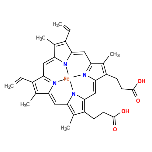
PROTOPORPHYRIN IX CONTAINING FE / Myeloperoxidase / Myeloperoxidase inhibitor SPIN
Similarity search - Component
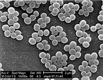
Biological speciesStaphylococcus aureus (bacteria)
Homo sapiens (human)
MethodX-RAY DIFFRACTION / SYNCHROTRON / MOLECULAR REPLACEMENT / Resolution: 2.55 Å
AuthorsLeitgeb, U. / Pfanzagl, V.
Funding support Austria, 2items
OrganizationGrant numberCountry
Austrian Science FundP33997 Austria
Austrian Science FundW1224 Austria
CitationJournal: Int J Biol Macromol / Year: 2026
Title: Interface N-glycans drive myeloperoxidase dimerization in vitro.
Authors: Urban Leitgeb / Yirui Guo / Tabitha Emde / Valentina Ruocco / Theodor Simak / Ela Zdenkovic / Paul G Furtmüller / Dominika Borek / William M Nauseef / Chris Oostenbrink / Vera Pfanzagl /
Abstract: Myeloperoxidase (MPO) is a highly glycosylated heme oxidoreductase that, together with lactoperoxidase (LPO) and eosinophil peroxidase (EPO), contributes to host defence through the generation of ...Myeloperoxidase (MPO) is a highly glycosylated heme oxidoreductase that, together with lactoperoxidase (LPO) and eosinophil peroxidase (EPO), contributes to host defence through the generation of (pseudo)hypohalous acids. Among the human heme peroxidases, mature MPO forms a unique covalently linked homodimer. Dimerization enhances stability and is essential for some biological functions, including chromatin disruption during neutrophil extracellular trap formation. Thus, we hypothesized that MPO dimerization must reflect a tightly regulated step in MPO biosynthesis and is driven by specific structural elements. Cross-species sequence alignment indicated that N-glycosylation motifs were evolutionarily more conserved in MPO than in monomeric EPO and LPO. We investigated the role of N-glycans in MPO dimerization using in vitro monomerized glycosylated (mMPO) and deglycosylated (mMPOdg) monomeric variants of native human MPO. Small-angle X-ray scattering, isothermal titration calorimetry and crystal structures showed that glycosylated MPO monomers had a weak but biologically relevant affinity in the millimolar range, which was lost upon deglycosylation. Furthermore, we present the first cryogenic transmission electron microscopy structure of human MPO with at least one core N-acetylglucosamine residue resolved at each N-glycosylation site. Structural analysis suggests that an extensive hydrogen bonding network of interface N-glycans at position 483 and residues of the other monomer drives MPO homodimerization. Dimerization enhances stability without affecting enzymatic activity. Together, we show that N-glycans are essential to MPO dimerization, provide important insights into the impact of interface N-glycans on biophysical and biochemical properties of MPO and discuss the implications for the biological roles of MPO in health and disease.
History
DepositionApr 10, 2026Deposition site: PDBE / Processing site: PDBE
Revision 1.0Oct 7, 2026Provider: repository / Type: Initial release

-
Structure visualization

Structure viewerMolecule:
MolmilJmol/JSmol

Downloads & links

-
Assembly

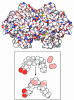
Deposited unit
A: Myeloperoxidase
B: Myeloperoxidase
E: Myeloperoxidase inhibitor SPIN
C: Myeloperoxidase
D: Myeloperoxidase
F: Myeloperoxidase inhibitor SPIN
hetero molecules


Theoretical massNumber of molelcules
Total (without water)155,03418
Polymers149,4736
Non-polymers5,56112
Water724
1
A: Myeloperoxidase
B: Myeloperoxidase
E: Myeloperoxidase inhibitor SPIN
hetero molecules


Theoretical massNumber of molelcules
Total (without water)77,3069
Polymers74,7083
Non-polymers2,5986
Water181
TypeNameSymmetry operationNumber
identity operation1_555x,y,z1
Buried area18080 Å2
ΔGint-79 kcal/mol
Surface area25300 Å2
2
C: Myeloperoxidase
D: Myeloperoxidase
F: Myeloperoxidase inhibitor SPIN
hetero molecules


Theoretical massNumber of molelcules
Total (without water)77,7289
Polymers74,7653
Non-polymers2,9636
Water181
TypeNameSymmetry operationNumber
identity operation1_555x,y,z1
Buried area18880 Å2
ΔGint-70 kcal/mol
Surface area25210 Å2
Unit cell
Length a, b, c (Å)111.474, 111.474, 241.912
Angle α, β, γ (deg.)90.00, 90.00, 90.00
Int Tables number96
Space group name H-MP43212

-
Components

-
Protein , 4 types, 6 molecules ACBEFD

#1: Protein Myeloperoxidase / MPO


Mass: 12951.516 Da / Num. of mol.: 2 / Source method: isolated from a natural source / Source: (natural) Homo sapiens (human) / References: UniProt: P05164, myeloperoxidase
#2: Protein Myeloperoxidase / MPO


Mass: 53362.316 Da / Num. of mol.: 1 / Source method: isolated from a natural source / Source: (natural) Homo sapiens (human) / References: UniProt: P05164, myeloperoxidase
#3: Protein Myeloperoxidase inhibitor SPIN


Mass: 8394.336 Da / Num. of mol.: 2
Source method: isolated from a genetically manipulated source
Source: (gene. exp.) Staphylococcus aureus (bacteria) / Gene: SAOUHSC_00401 / Production host: Escherichia coli (E. coli) / References: UniProt: Q2G0X2
#4: Protein Myeloperoxidase / MPO


Mass: 53419.367 Da / Num. of mol.: 1 / Source method: isolated from a natural source / Source: (natural) Homo sapiens (human) / References: UniProt: P05164, myeloperoxidase

-
Sugars , 4 types, 6 molecules

#5: Polysaccharide alpha-D-mannopyranose-(1-3)-[alpha-D-mannopyranose-(1-6)]beta-D-mannopyranose-(1-4)-2-acetamido-2- ...alpha-D-mannopyranose-(1-3)-[alpha-D-mannopyranose-(1-6)]beta-D-mannopyranose-(1-4)-2-acetamido-2-deoxy-beta-D-glucopyranose-(1-4)-[alpha-L-fucopyranose-(1-6)]2-acetamido-2-deoxy-beta-D-glucopyranose


Type: oligosaccharide / Mass: 1056.964 Da / Num. of mol.: 1
Source method: isolated from a genetically manipulated source
DescriptorTypeProgram
DManpa1-3[DManpa1-6]DManpb1-4DGlcpNAcb1-4[LFucpa1-6]DGlcpNAcb1-ROHGlycam Condensed SequenceGMML 1.0
WURCS=2.0/4,6,5/[a2122h-1b_1-5_2*NCC/3=O][a1122h-1b_1-5][a1122h-1a_1-5][a1221m-1a_1-5]/1-1-2-3-3-4/a4-b1_a6-f1_b4-c1_c3-d1_c6-e1WURCSPDB2Glycan 1.1.0
[][D-1-deoxy-GlcpNAc]{[(4+1)][b-D-GlcpNAc]{[(4+1)][b-D-Manp]{[(3+1)][a-D-Manp]{}[(6+1)][a-D-Manp]{}}}[(6+1)][a-L-Fucp]{}}LINUCSPDB-CARE
#6: Polysaccharide 2-acetamido-2-deoxy-beta-D-glucopyranose-(1-4)-2-acetamido-2-deoxy-beta-D-glucopyranose


Type: oligosaccharide / Mass: 424.401 Da / Num. of mol.: 3
Source method: isolated from a genetically manipulated source
DescriptorTypeProgram
DGlcpNAcb1-4DGlcpNAcb1-ROHGlycam Condensed SequenceGMML 1.0
WURCS=2.0/1,2,1/[a2122h-1b_1-5_2*NCC/3=O]/1-1/a4-b1WURCSPDB2Glycan 1.1.0
[][D-1-deoxy-GlcpNAc]{[(4+1)][b-D-GlcpNAc]{}}LINUCSPDB-CARE
#7: Polysaccharide 2-acetamido-2-deoxy-beta-D-glucopyranose-(1-2)-alpha-D-mannopyranose-(1-6)-[alpha-D-mannopyranose- ...2-acetamido-2-deoxy-beta-D-glucopyranose-(1-2)-alpha-D-mannopyranose-(1-6)-[alpha-D-mannopyranose-(1-3)]beta-D-mannopyranose-(1-4)-2-acetamido-2-deoxy-beta-D-glucopyranose-(1-4)-[alpha-L-fucopyranose-(1-6)]2-acetamido-2-deoxy-beta-D-glucopyranose


Type: oligosaccharide / Mass: 1260.157 Da / Num. of mol.: 1
Source method: isolated from a genetically manipulated source
DescriptorTypeProgram
DGlcpNAcb1-2DManpa1-6[DManpa1-3]DManpb1-4DGlcpNAcb1-4[LFucpa1-6]DGlcpNAcb1-ROHGlycam Condensed SequenceGMML 1.0
WURCS=2.0/4,7,6/[a2122h-1b_1-5_2*NCC/3=O][a1122h-1b_1-5][a1122h-1a_1-5][a1221m-1a_1-5]/1-1-2-3-3-1-4/a4-b1_a6-g1_b4-c1_c3-d1_c6-e1_e2-f1WURCSPDB2Glycan 1.1.0
[][D-1-deoxy-GlcpNAc]{[(4+1)][b-D-GlcpNAc]{[(4+1)][b-D-Manp]{[(3+1)][a-D-Manp]{}[(6+1)][a-D-Manp]{[(2+1)][b-D-GlcpNAc]{}}}}[(6+1)][a-L-Fucp]{}}LINUCSPDB-CARE
#8: Polysaccharide beta-D-mannopyranose-(1-4)-2-acetamido-2-deoxy-beta-D-glucopyranose-(1-4)-2-acetamido-2-deoxy-beta- ...beta-D-mannopyranose-(1-4)-2-acetamido-2-deoxy-beta-D-glucopyranose-(1-4)-2-acetamido-2-deoxy-beta-D-glucopyranose


Type: oligosaccharide / Mass: 586.542 Da / Num. of mol.: 1
Source method: isolated from a genetically manipulated source
DescriptorTypeProgram
DManpb1-4DGlcpNAcb1-4DGlcpNAcb1-ROHGlycam Condensed SequenceGMML 1.0
WURCS=2.0/2,3,2/[a2122h-1b_1-5_2*NCC/3=O][a1122h-1b_1-5]/1-1-2/a4-b1_b4-c1WURCSPDB2Glycan 1.1.0
[][D-1-deoxy-GlcpNAc]{[(4+1)][b-D-GlcpNAc]{[(4+1)][b-D-Manp]{}}}LINUCSPDB-CARE

-
Non-polymers , 4 types, 10 molecules

#9: Chemical ChemComp-CL / CHLORIDE ION


Mass: 35.453 Da / Num. of mol.: 2 / Source method: obtained synthetically / Formula: Cl
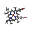
#10: Chemical ChemComp-HEM / PROTOPORPHYRIN IX CONTAINING FE / HEME


Mass: 616.487 Da / Num. of mol.: 2 / Source method: obtained synthetically / Formula: C34H32FeN4O4 / Feature type: SUBJECT OF INVESTIGATION
#11: Chemical ChemComp-CA / CALCIUM ION


Mass: 40.078 Da / Num. of mol.: 2 / Source method: obtained synthetically / Formula: Ca
#12: Water ChemComp-HOH / water


Mass: 18.015 Da / Num. of mol.: 4 / Source method: isolated from a natural source / Formula: H2O

-
Details

Has ligand of interestY
Has protein modificationY

-
Experimental details

-
Experiment

ExperimentMethod: X-RAY DIFFRACTION / Number of used crystals: 1

-
Sample preparation

CrystalDensity Matthews: 2.52 Å3/Da / Density % sol: 51.29 %
Crystal growTemperature: 293.15 K / Method: vapor diffusion, sitting drop / pH: 7.5
Details: 0.1 M TRIS 7.5 pH, 8 %w/v PEG 1K, 8 %w/v PEG 8K, 0.4 M KSCN

-
Data collection

DiffractionMean temperature: 213.15 K / Serial crystal experiment: N
Diffraction sourceSource: SYNCHROTRON / Site: ESRF / Beamline: MASSIF-3 / Wavelength: 0.9677 Å
DetectorType: DECTRIS EIGER X 4M / Detector: PIXEL / Date: Nov 3, 2023
RadiationMonochromator: Si(111) / Protocol: SINGLE WAVELENGTH / Monochromatic (M) / Laue (L): M / Scattering type: x-ray
Radiation wavelengthWavelength: 0.9677 Å / Relative weight: 1
ReflectionResolution: 2.55→78.95 Å / Num. obs: 30405 / % possible obs: 95 % / Redundancy: 12.4 % / CC1/2: 0.995 / Rmerge(I) obs: 0.21 / Rpim(I) all: 0.062 / Rrim(I) all: 0.219 / Net I/σ(I): 8.4
Reflection shellResolution: 2.55→2.88 Å / Redundancy: 12.3 % / Rmerge(I) obs: 1.468 / Mean I/σ(I) obs: 1.6 / Num. unique obs: 1520 / CC1/2: 0.714 / Rpim(I) all: 0.433 / Rrim(I) all: 1.532 / % possible all: 73.5

-
Processing

Software
NameVersionClassification
REFMAC5.8.0431refinement
Aimlessdata scaling
XDSdata reduction
MOLREPphasing
PDB_EXTRACTdata extraction
RefinementMethod to determine structure: MOLECULAR REPLACEMENT / Resolution: 2.55→78.95 Å / Cor.coef. Fo:Fc: 0.92 / Cor.coef. Fo:Fc free: 0.861 / SU B: 34.639 / SU ML: 0.335 / Cross valid method: THROUGHOUT / ESU R Free: 0.49 / Stereochemistry target values: MAXIMUM LIKELIHOOD / Details: HYDROGENS HAVE BEEN USED IF PRESENT IN THE INPUT
RfactorNum. reflection% reflectionSelection details
Rfree0.26438 1509 5 %RANDOM
Rwork0.2017 ---
obs0.20485 28896 60.25 %-
Solvent computationIon probe radii: 0.8 Å / Shrinkage radii: 0.8 Å / VDW probe radii: 1.2 Å / Solvent model: MASK
Displacement parametersBiso mean: 61.293 Å2
Baniso -1Baniso -2Baniso -3
1--0.71 Å20 Å20 Å2
2---0.71 Å20 Å2
3---1.42 Å2
Refinement stepCycle: 1 / Resolution: 2.55→78.95 Å
ProteinNucleic acidLigandSolventTotal
Num. atoms10150 0 369 4 10523
Refine LS restraints
Refine-IDTypeDev idealDev ideal targetNumber
X-RAY DIFFRACTIONr_bond_refined_d0.0060.01210803
X-RAY DIFFRACTIONr_bond_other_d0.0010.01610070
X-RAY DIFFRACTIONr_angle_refined_deg1.4791.84714715
X-RAY DIFFRACTIONr_angle_other_deg0.4961.76723167
X-RAY DIFFRACTIONr_dihedral_angle_1_deg7.23151255
X-RAY DIFFRACTIONr_dihedral_angle_2_deg10.3215130
X-RAY DIFFRACTIONr_dihedral_angle_3_deg13.74101757
X-RAY DIFFRACTIONr_dihedral_angle_4_deg
X-RAY DIFFRACTIONr_chiral_restr0.0640.21619
X-RAY DIFFRACTIONr_gen_planes_refined0.0060.0212932
X-RAY DIFFRACTIONr_gen_planes_other0.0010.022640
X-RAY DIFFRACTIONr_nbd_refined
X-RAY DIFFRACTIONr_nbd_other
X-RAY DIFFRACTIONr_nbtor_refined
X-RAY DIFFRACTIONr_nbtor_other
X-RAY DIFFRACTIONr_xyhbond_nbd_refined
X-RAY DIFFRACTIONr_xyhbond_nbd_other
X-RAY DIFFRACTIONr_metal_ion_refined
X-RAY DIFFRACTIONr_metal_ion_other
X-RAY DIFFRACTIONr_symmetry_vdw_refined
X-RAY DIFFRACTIONr_symmetry_vdw_other
X-RAY DIFFRACTIONr_symmetry_hbond_refined
X-RAY DIFFRACTIONr_symmetry_hbond_other
X-RAY DIFFRACTIONr_symmetry_metal_ion_refined
X-RAY DIFFRACTIONr_symmetry_metal_ion_other
X-RAY DIFFRACTIONr_mcbond_it1.6862.4765038
X-RAY DIFFRACTIONr_mcbond_other1.6862.4765038
X-RAY DIFFRACTIONr_mcangle_it2.8784.4476287
X-RAY DIFFRACTIONr_mcangle_other2.8774.4476288
X-RAY DIFFRACTIONr_scbond_it2.1632.6785765
X-RAY DIFFRACTIONr_scbond_other2.1632.6785764
X-RAY DIFFRACTIONr_scangle_it
X-RAY DIFFRACTIONr_scangle_other3.4634.8818429
X-RAY DIFFRACTIONr_long_range_B_refined6.33330.1442671
X-RAY DIFFRACTIONr_long_range_B_other6.33330.1442672
X-RAY DIFFRACTIONr_rigid_bond_restr
X-RAY DIFFRACTIONr_sphericity_free
X-RAY DIFFRACTIONr_sphericity_bonded
LS refinement shellResolution: 2.552→2.618 Å / Total num. of bins used: 20
RfactorNum. reflection% reflection
Rfree0 0 -
Rwork0.404 44 -
obs--1.21 %
Refinement TLS params.

Method: refined / Refine-ID: X-RAY DIFFRACTION

IDL11 (°2)L12 (°2)L13 (°2)L22 (°2)L23 (°2)L33 (°2)S11 (Å °)S12 (Å °)S13 (Å °)S21 (Å °)S22 (Å °)S23 (Å °)S31 (Å °)S32 (Å °)S33 (Å °)T11 (Å2)T12 (Å2)T13 (Å2)T22 (Å2)T23 (Å2)T33 (Å2)Origin x (Å)Origin y (Å)Origin z (Å)
11.50220.27340.10012.55340.28451.73390.3443-0.07920.14060.0017-0.26370.19770.0007-0.127-0.08060.16120.08950.04420.1696-0.00670.04-9.58715.858921.6619
21.44970.3124-0.04172.1616-0.22911.47760.3372-0.0253-0.1431-0.0758-0.32290.01420.1612-0.043-0.01430.21970.1061-0.00940.1780.00260.0303-6.82824.516822.927
31.8326-1.3098-0.16131.1070.33734.10880.0638-0.1306-0.4480.2323-0.0670.3772-0.2009-0.12480.00320.5681-0.15930.02460.6073-0.05840.468-27.6047-5.686943.1547
41.8386-0.0358-0.02752.952-0.10021.56130.2383-0.16790.33480.2461-0.2738-0.0632-0.0781-0.06610.03540.2264-0.12610.20770.1866-0.11790.2336-3.361742.041343.7219
51.3713-0.40360.47192.211-0.1781.27580.1437-0.0690.56880.2465-0.1943-0.0563-0.3933-0.06120.05060.3259-0.10160.24510.2605-0.09960.4258-2.03153.650640.8722
60.5766-0.03181.38690.109-0.21683.58820.00150.01570.2421-0.1457-0.01550.305-0.04370.10120.0140.92130.06270.08961.0824-0.02841.4289-30.50463.468735.3266
Refinement TLS group
IDRefine-IDRefine TLS-IDAuth asym-IDAuth seq-ID
1X-RAY DIFFRACTION1A171 - 271
2X-RAY DIFFRACTION2B280 - 759
3X-RAY DIFFRACTION3E30 - 98
4X-RAY DIFFRACTION4C172 - 269
5X-RAY DIFFRACTION5D287 - 763
6X-RAY DIFFRACTION6F30 - 99

+
About Yorodumi

-
News

-
Feb 9, 2022. New format data for meta-information of EMDB entries

New format data for meta-information of EMDB entries

  • Version 3 of the EMDB header file is now the official format.
  • The previous official version 1.9 will be removed from the archive.

Related info.:EMDB header

External links:wwPDB to switch to version 3 of the EMDB data model

-
Aug 12, 2020. Covid-19 info

Covid-19 info

URL: https://pdbj.org/emnavi/covid19.php

New page: Covid-19 featured information page in EM Navigator.

Related info.:Covid-19 info / Mar 5, 2020. Novel coronavirus structure data

+
Mar 5, 2020. Novel coronavirus structure data

Novel coronavirus structure data

Related info.:Yorodumi Speices / Aug 12, 2020. Covid-19 info

External links:COVID-19 featured content - PDBj / Molecule of the Month (242):Coronavirus Proteases

+
Jan 31, 2019. EMDB accession codes are about to change! (news from PDBe EMDB page)

EMDB accession codes are about to change! (news from PDBe EMDB page)

  • The allocation of 4 digits for EMDB accession codes will soon come to an end. Whilst these codes will remain in use, new EMDB accession codes will include an additional digit and will expand incrementally as the available range of codes is exhausted. The current 4-digit format prefixed with “EMD-” (i.e. EMD-XXXX) will advance to a 5-digit format (i.e. EMD-XXXXX), and so on. It is currently estimated that the 4-digit codes will be depleted around Spring 2019, at which point the 5-digit format will come into force.
  • The EM Navigator/Yorodumi systems omit the EMD- prefix.

Related info.:Q: What is EMD? / ID/Accession-code notation in Yorodumi/EM Navigator

External links:EMDB Accession Codes are Changing Soon! / Contact to PDBj

+
Jul 12, 2017. Major update of PDB

Major update of PDB

  • wwPDB released updated PDB data conforming to the new PDBx/mmCIF dictionary.
  • This is a major update changing the version number from 4 to 5, and with Remediation, in which all the entries are updated.
  • In this update, many items about electron microscopy experimental information are reorganized (e.g. em_software).
  • Now, EM Navigator and Yorodumi are based on the updated data.

External links:wwPDB Remediation / Enriched Model Files Conforming to OneDep Data Standards Now Available in the PDB FTP Archive

-
Yorodumi

Thousand views of thousand structures

  • Yorodumi is a browser for structure data from EMDB, PDB, SASBDB, etc.
  • This page is also the successor to EM Navigator detail page, and also detail information page/front-end page for Omokage search.
  • The word "yorodu" (or yorozu) is an old Japanese word meaning "ten thousand". "mi" (miru) is to see.

Related info.:EMDB / PDB / SASBDB / Comparison of 3 databanks / Yorodumi Search / Aug 31, 2016. New EM Navigator & Yorodumi / Yorodumi Papers / Jmol/JSmol / Function and homology information / Changes in new EM Navigator and Yorodumi

Read more