 Movie
Movie Controller

[English] 日本語
 Yorodumi
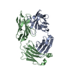
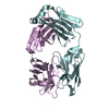
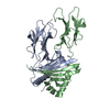
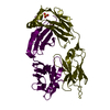
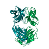
- PDB-2zz8: Crystal structure of LipL32, the most abundant surface protein of... -
+ Open data

- Basic information
| Entry | Database: PDB / ID: 2zz8 | ||||||
|---|---|---|---|---|---|---|---|
| Title | Crystal structure of LipL32, the most abundant surface protein of pathogenic leptospira spp | ||||||
 Components Components | LipL32 protein | ||||||
 Keywords Keywords | UNKNOWN FUNCTION / Leptospira / outer-membrane protein | ||||||
| Function / homology | Surface lipoprotein of Spirochaetales order / Surface lipoprotein of Spirochaetales order / Prokaryotic membrane lipoprotein lipid attachment site profile. / LipL32 Function and homology information Function and homology information | ||||||
| Biological species |  Leptospira interrogans (bacteria) Leptospira interrogans (bacteria) | ||||||
| Method |  X-RAY DIFFRACTION / SYNCHROTRON / SAD / Resolution: 2.01 Å X-RAY DIFFRACTION / SYNCHROTRON / SAD / Resolution: 2.01 Å | ||||||
 Authors Authors | Vivian, J.P. / Beddoe, T. / Rossjohn, J. | ||||||
 Citation Citation | Journal: J.Mol.Biol. / Year: 2009 Title: Crystal structure of LipL32, the most abundant surface protein of pathogenic Leptospira spp. Authors: Vivian, J.P. / Beddoe, T. / McAlister, A.D. / Wilce, M.C. / Zaker-Tabrizi, L. / Troy, S. / Byres, E. / Hoke, D.E. / Cullen, P.A. / Lo, M. / Murray, G.L. / Adler, B. / Rossjohn, J. | ||||||
| History |
|
- Structure visualization
| Structure viewer | Molecule: MolmilJmol/JSmol |
|---|
- Downloads & links
-Download
| PDBx/mmCIF format | 2zz8.cif.gz | 108.7 KB | Display | PDBx/mmCIF format |
|---|---|---|---|---|
| PDB format | pdb2zz8.ent.gz | 84.9 KB | Display | PDB format |
| PDBx/mmJSON format | 2zz8.json.gz | Tree view | PDBx/mmJSON format | |
| Others |  Other downloads Other downloads |
-Validation report
| Summary document | 2zz8_validation.pdf.gz | 441.8 KB | Display | wwPDB validaton report |
|---|---|---|---|---|
| Full document | 2zz8_full_validation.pdf.gz | 450.8 KB | Display | |
| Data in XML | 2zz8_validation.xml.gz | 24 KB | Display | |
| Data in CIF | 2zz8_validation.cif.gz | 35.1 KB | Display | |
| Arichive directory | https://data.pdbj.org/pub/pdb/validation_reports/zz/2zz8ftp://data.pdbj.org/pub/pdb/validation_reports/zz/2zz8 | HTTPS FTP |
-Related structure data
| Similar structure data |
|---|


-Links
 PDBj
PDBj


- Assembly
| Deposited unit | 
| ||||||||
|---|---|---|---|---|---|---|---|---|---|
| 1 |
| ||||||||
| Unit cell |
|
-Components
| #1: Protein | Mass: 26480.059 Da / Num. of mol.: 2 / Fragment: residues in database 21-261 Source method: isolated from a genetically manipulated source Source: (gene. exp.) Leptospira interrogans (bacteria) / Strain: Lai / Gene: lipl32 / Plasmid: pQE30 / Production host: #2: Water | ChemComp-HOH / |  Mass: 18.015 Da / Num. of mol.: 435 / Source method: isolated from a natural source / Formula: H2O Mass: 18.015 Da / Num. of mol.: 435 / Source method: isolated from a natural source / Formula: H2O |
|---|
-Experimental details
-Experiment
| Experiment | Method: X-RAY DIFFRACTION / Number of used crystals: 2 |
|---|
- Sample preparation
| Crystal | Density Matthews: 3.97 Å3/Da / Density % sol: 68.98 % |
|---|---|
| Crystal grow | Temperature: 294 K / Method: vapor diffusion, hanging drop / pH: 6.2 Details: 2.0M sodium malonate, 0.1M sodium cacodylate, 4%(v/v) butyrolactone, pH 6.2, VAPOR DIFFUSION, HANGING DROP, temperature 294K |
-Data collection
| Diffraction |
| ||||||||||||||||||
|---|---|---|---|---|---|---|---|---|---|---|---|---|---|---|---|---|---|---|---|
| Diffraction source |
| ||||||||||||||||||
| Detector |
| ||||||||||||||||||
| Radiation |
| ||||||||||||||||||
| Radiation wavelength |
| ||||||||||||||||||
| Reflection | Resolution: 2→40 Å / Num. all: 59453 / Num. obs: 59453 / % possible obs: 100 % / Observed criterion σ(F): 1 / Observed criterion σ(I): 1 / Redundancy: 21.4 % / Biso Wilson estimate: 38 Å2 / Rmerge(I) obs: 0.078 / Rsym value: 0.08 / Net I/σ(I): 41.9 | ||||||||||||||||||
| Reflection shell | Resolution: 2→2.11 Å / Redundancy: 20.3 % / Rmerge(I) obs: 0.72 / Mean I/σ(I) obs: 2.4 / % possible all: 100 |

- Processing
| Software |
| ||||||||||||||||||||||||||||||||||||||||||||||||||||||||||||||||||||||||||||||||||||||||||||||||||||||||||||||||||||||||||||||||||||||||||||||||||||||||||||||||||||||||||
|---|---|---|---|---|---|---|---|---|---|---|---|---|---|---|---|---|---|---|---|---|---|---|---|---|---|---|---|---|---|---|---|---|---|---|---|---|---|---|---|---|---|---|---|---|---|---|---|---|---|---|---|---|---|---|---|---|---|---|---|---|---|---|---|---|---|---|---|---|---|---|---|---|---|---|---|---|---|---|---|---|---|---|---|---|---|---|---|---|---|---|---|---|---|---|---|---|---|---|---|---|---|---|---|---|---|---|---|---|---|---|---|---|---|---|---|---|---|---|---|---|---|---|---|---|---|---|---|---|---|---|---|---|---|---|---|---|---|---|---|---|---|---|---|---|---|---|---|---|---|---|---|---|---|---|---|---|---|---|---|---|---|---|---|---|---|---|---|---|---|---|---|
| Refinement | Method to determine structure: SAD / Resolution: 2.01→37.88 Å / Cor.coef. Fo:Fc: 0.968 / Cor.coef. Fo:Fc free: 0.955 / SU B: 7.207 / SU ML: 0.088 / TLS residual ADP flag: LIKELY RESIDUAL / Cross valid method: THROUGHOUT / ESU R: 0.117 / ESU R Free: 0.118 / Stereochemistry target values: MAXIMUM LIKELIHOOD
| ||||||||||||||||||||||||||||||||||||||||||||||||||||||||||||||||||||||||||||||||||||||||||||||||||||||||||||||||||||||||||||||||||||||||||||||||||||||||||||||||||||||||||
| Solvent computation | Ion probe radii: 0.8 Å / Shrinkage radii: 0.8 Å / VDW probe radii: 1.2 Å / Solvent model: MASK | ||||||||||||||||||||||||||||||||||||||||||||||||||||||||||||||||||||||||||||||||||||||||||||||||||||||||||||||||||||||||||||||||||||||||||||||||||||||||||||||||||||||||||
| Displacement parameters | Biso mean: 25.374 Å2
| ||||||||||||||||||||||||||||||||||||||||||||||||||||||||||||||||||||||||||||||||||||||||||||||||||||||||||||||||||||||||||||||||||||||||||||||||||||||||||||||||||||||||||
| Refinement step | Cycle: LAST / Resolution: 2.01→37.88 Å
| ||||||||||||||||||||||||||||||||||||||||||||||||||||||||||||||||||||||||||||||||||||||||||||||||||||||||||||||||||||||||||||||||||||||||||||||||||||||||||||||||||||||||||
| Refine LS restraints |
| ||||||||||||||||||||||||||||||||||||||||||||||||||||||||||||||||||||||||||||||||||||||||||||||||||||||||||||||||||||||||||||||||||||||||||||||||||||||||||||||||||||||||||
| LS refinement shell | Resolution: 2.006→2.058 Å / Total num. of bins used: 20
| ||||||||||||||||||||||||||||||||||||||||||||||||||||||||||||||||||||||||||||||||||||||||||||||||||||||||||||||||||||||||||||||||||||||||||||||||||||||||||||||||||||||||||
| Refinement TLS params. | Method: refined / Refine-ID: X-RAY DIFFRACTION
| ||||||||||||||||||||||||||||||||||||||||||||||||||||||||||||||||||||||||||||||||||||||||||||||||||||||||||||||||||||||||||||||||||||||||||||||||||||||||||||||||||||||||||
| Refinement TLS group |
|
