 Movie
Movie Controller

[English] 日本語
 Yorodumi
- PDB-2zw2: Crystal Structure of Formylglycinamide Ribonucleotide Amidotransf... -
+ Open data

- Basic information
| Entry | Database: PDB / ID: 2zw2 | ||||||
|---|---|---|---|---|---|---|---|
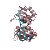
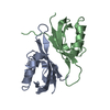
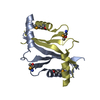
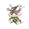
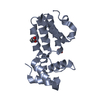
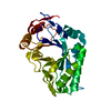
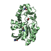
| Title | Crystal Structure of Formylglycinamide Ribonucleotide Amidotransferase III from SULFOLOBUS TOKODAII (STPURS) | ||||||
 Components Components | Putative uncharacterized protein STS178 | ||||||
 Keywords Keywords | LIGASE / Purine metabolism | ||||||
| Function / homology |  Function and homology information Function and homology informationphosphoribosylformylglycinamidine synthase / phosphoribosylformylglycinamidine synthase activity / 'de novo' IMP biosynthetic process / ATP binding / cytoplasm Similarity search - Function | ||||||
| Biological species |   Sulfolobus tokodaii (archaea) Sulfolobus tokodaii (archaea) | ||||||
| Method |  X-RAY DIFFRACTION / SYNCHROTRON / MOLECULAR REPLACEMENT / Resolution: 1.55 Å X-RAY DIFFRACTION / SYNCHROTRON / MOLECULAR REPLACEMENT / Resolution: 1.55 Å | ||||||
 Authors Authors | Suzuki, S. / Tamura, S. / Okada, K. / Baba, S. / Kumasaka, T. / Nakagawa, N. / Masui, R. / Kuramitsu, S. / Sampei, G. / Kawai, G. | ||||||
 Citation Citation | Journal: To be Published Title: Crystal Structure of Formylglycinamide Ribonucleotide Amidotransferase III from SULFOLOBUS TOKODAII (STPURS) Authors: Suzuki, S. / Tamura, S. / Okada, K. / Baba, S. / Kumasaka, T. / Nakagawa, N. / Masui, R. / Kuramitsu, S. / Sampei, G. / Kawai, G. | ||||||
| History |
|
- Structure visualization
| Structure viewer | Molecule: MolmilJmol/JSmol |
|---|
- Downloads & links
-Download
| PDBx/mmCIF format | 2zw2.cif.gz | 51.4 KB | Display | PDBx/mmCIF format |
|---|---|---|---|---|
| PDB format | pdb2zw2.ent.gz | 36.8 KB | Display | PDB format |
| PDBx/mmJSON format | 2zw2.json.gz | Tree view | PDBx/mmJSON format | |
| Others |  Other downloads Other downloads |
-Validation report
| Arichive directory | https://data.pdbj.org/pub/pdb/validation_reports/zw/2zw2ftp://data.pdbj.org/pub/pdb/validation_reports/zw/2zw2 | HTTPS FTP |
|---|
-Related structure data
| Related structure data | 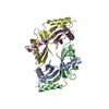 1vq3S S: Starting model for refinement |
|---|---|
| Similar structure data |


-Links
 PDBj
PDBj- Assembly
| Deposited unit | 
| ||||||||
|---|---|---|---|---|---|---|---|---|---|
| 1 |
| ||||||||
| Unit cell |
|
-Components
| #1: Protein | Mass: 10763.329 Da / Num. of mol.: 2 Source method: isolated from a genetically manipulated source Source: (gene. exp.) Sulfolobus tokodaii (archaea) / Strain: 7 / Gene: purS / Plasmid: pET-11a / Production host:  References: UniProt: Q970V8, UniProt: F9VNF4*PLUS, phosphoribosylformylglycinamidine synthase #2: Chemical |   Mass: 92.094 Da / Num. of mol.: 2 / Source method: obtained synthetically / Formula: C3H8O3 Mass: 92.094 Da / Num. of mol.: 2 / Source method: obtained synthetically / Formula: C3H8O3#3: Water | ChemComp-HOH / |  Mass: 18.015 Da / Num. of mol.: 200 / Source method: isolated from a natural source / Formula: H2O Mass: 18.015 Da / Num. of mol.: 200 / Source method: isolated from a natural source / Formula: H2O |
|---|
-Experimental details
-Experiment
| Experiment | Method: X-RAY DIFFRACTION / Number of used crystals: 1 |
|---|
- Sample preparation
| Crystal | Density Matthews: 1.95 Å3/Da / Density % sol: 36.8 % |
|---|---|
| Crystal grow | Temperature: 293 K / Method: vapor diffusion, hanging drop / pH: 7.5 Details: 0.1M HEPES sodium pH7.5, 0.8M potassium sodium tartrate tetrahydrate, VAPOR DIFFUSION, HANGING DROP, temperature 293.0K |
-Data collection
| Diffraction | Mean temperature: 100 K |
|---|---|
| Diffraction source | Source: SYNCHROTRON / Site: SPring-8  / Beamline: BL38B1 / Wavelength: 1 Å / Beamline: BL38B1 / Wavelength: 1 Å |
| Detector | Type: RIGAKU JUPITER 210 / Detector: CCD / Date: Jun 30, 2008 |
| Radiation | Monochromator: Fixed exit Si 111 double crystal monochromator Protocol: SINGLE WAVELENGTH / Monochromatic (M) / Laue (L): M / Scattering type: x-ray |
| Radiation wavelength | Wavelength: 1 Å / Relative weight: 1 |
| Reflection | Resolution: 1.55→50 Å / Num. all: 24008 / Num. obs: 24008 / % possible obs: 97.3 % / Redundancy: 6.9 % / Biso Wilson estimate: 21.4 Å2 / Rmerge(I) obs: 0.03 / Net I/σ(I): 17.4 |
| Reflection shell | Resolution: 1.55→1.61 Å / Redundancy: 4.9 % / Rmerge(I) obs: 0.329 / Mean I/σ(I) obs: 3.3 / Num. unique all: 1925 / % possible all: 79.7 |
- Processing
| Software |
| ||||||||||||||||||||||||||||||||||||||||||||||||||||||||||||
|---|---|---|---|---|---|---|---|---|---|---|---|---|---|---|---|---|---|---|---|---|---|---|---|---|---|---|---|---|---|---|---|---|---|---|---|---|---|---|---|---|---|---|---|---|---|---|---|---|---|---|---|---|---|---|---|---|---|---|---|---|---|
| Refinement | Method to determine structure: MOLECULAR REPLACEMENT Starting model: PDB ENTRY 1VQ3 Resolution: 1.55→24.74 Å / Rfactor Rfree error: 0.005 / Data cutoff high absF: 315431.83 / Data cutoff low absF: 0 / Isotropic thermal model: RESTRAINED / Cross valid method: THROUGHOUT / σ(F): 0
| ||||||||||||||||||||||||||||||||||||||||||||||||||||||||||||
| Solvent computation | Solvent model: FLAT MODEL / Bsol: 65.154 Å2 / ksol: 0.408065 e/Å3 | ||||||||||||||||||||||||||||||||||||||||||||||||||||||||||||
| Displacement parameters | Biso mean: 21.9 Å2
| ||||||||||||||||||||||||||||||||||||||||||||||||||||||||||||
| Refine analyze |
| ||||||||||||||||||||||||||||||||||||||||||||||||||||||||||||
| Refinement step | Cycle: LAST / Resolution: 1.55→24.74 Å
| ||||||||||||||||||||||||||||||||||||||||||||||||||||||||||||
| Refine LS restraints |
| ||||||||||||||||||||||||||||||||||||||||||||||||||||||||||||
| LS refinement shell | Resolution: 1.55→1.65 Å / Rfactor Rfree error: 0.017 / Total num. of bins used: 6
| ||||||||||||||||||||||||||||||||||||||||||||||||||||||||||||
| Xplor file |
|
