 Movie
Movie Controller

[English] 日本語
 Yorodumi
- PDB-2zux: Crystal structure of rhamnogalacturonan lyase YesW complexed with... -
+ Open data

- Basic information
| Entry | Database: PDB / ID: 2zux | ||||||
|---|---|---|---|---|---|---|---|
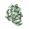
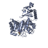
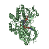
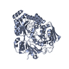
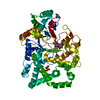
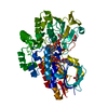
| Title | Crystal structure of rhamnogalacturonan lyase YesW complexed with rhamnose | ||||||
 Components Components | YesW protein | ||||||
 Keywords Keywords | LYASE / Beta-propeller / Rhamnose complex | ||||||
| Function / homology |  Function and homology information Function and homology informationrhamnogalacturonan endolyase / rhamnogalacturonan endolyase activity / cell wall organization / extracellular region / metal ion binding Similarity search - Function | ||||||
| Biological species |  | ||||||
| Method |  X-RAY DIFFRACTION / SYNCHROTRON / MOLECULAR REPLACEMENT / Resolution: 1.32 Å X-RAY DIFFRACTION / SYNCHROTRON / MOLECULAR REPLACEMENT / Resolution: 1.32 Å | ||||||
 Authors Authors | Ochiai, A. / Itoh, T. / Mikami, B. / Hashimoto, W. / Murata, K. | ||||||
 Citation Citation | Journal: J.Biol.Chem. / Year: 2009 Title: Structural determinants responsible for substrate recognition and mode of action in family 11 polysaccharide lyases Authors: Ochiai, A. / Itoh, T. / Mikami, B. / Hashimoto, W. / Murata, K. | ||||||
| History |
|
- Structure visualization
| Structure viewer | Molecule: MolmilJmol/JSmol |
|---|
- Downloads & links
-Download
| PDBx/mmCIF format | 2zux.cif.gz | 268.7 KB | Display | PDBx/mmCIF format |
|---|---|---|---|---|
| PDB format | pdb2zux.ent.gz | 211.7 KB | Display | PDB format |
| PDBx/mmJSON format | 2zux.json.gz | Tree view | PDBx/mmJSON format | |
| Others |  Other downloads Other downloads |
-Validation report
| Summary document | 2zux_validation.pdf.gz | 473.5 KB | Display | wwPDB validaton report |
|---|---|---|---|---|
| Full document | 2zux_full_validation.pdf.gz | 484.6 KB | Display | |
| Data in XML | 2zux_validation.xml.gz | 54.4 KB | Display | |
| Data in CIF | 2zux_validation.cif.gz | 83.3 KB | Display | |
| Arichive directory | https://data.pdbj.org/pub/pdb/validation_reports/zu/2zuxftp://data.pdbj.org/pub/pdb/validation_reports/zu/2zux | HTTPS FTP |
-Related structure data
| Related structure data |  2zuyC  2z8rS C: citing same article ( S: Starting model for refinement |
|---|---|
| Similar structure data |

-Links
 PDBj
PDBj
- Assembly
| Deposited unit | 
| ||||||||
|---|---|---|---|---|---|---|---|---|---|
| 1 | 
| ||||||||
| 2 | 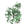
| ||||||||
| Unit cell |
|
-Components
| #1: Protein | Mass: 64520.133 Da / Num. of mol.: 2 / Fragment: UNP residues 38-620 Source method: isolated from a genetically manipulated source Source: (gene. exp.) References: UniProt: O31526, Lyases; Carbon-oxygen lyases; Acting on polysaccharides #2: Chemical | ChemComp-CA /   Mass: 40.078 Da / Num. of mol.: 20 / Source method: obtained synthetically / Formula: Ca Mass: 40.078 Da / Num. of mol.: 20 / Source method: obtained synthetically / Formula: Ca#3: Sugar | ChemComp-RAM /   Type: L-saccharide, alpha linking / Mass: 164.156 Da / Num. of mol.: 7 Type: L-saccharide, alpha linking / Mass: 164.156 Da / Num. of mol.: 7Source method: isolated from a genetically manipulated source Formula: C6H12O5 #4: Chemical |   Mass: 118.174 Da / Num. of mol.: 2 / Source method: obtained synthetically / Formula: C6H14O2 / Comment: precipitant*YM Mass: 118.174 Da / Num. of mol.: 2 / Source method: obtained synthetically / Formula: C6H14O2 / Comment: precipitant*YM#5: Water | ChemComp-HOH / |  Mass: 18.015 Da / Num. of mol.: 1255 / Source method: isolated from a natural source / Formula: H2O Mass: 18.015 Da / Num. of mol.: 1255 / Source method: isolated from a natural source / Formula: H2O |
|---|
-Experimental details
-Experiment
| Experiment | Method: X-RAY DIFFRACTION / Number of used crystals: 1 |
|---|
- Sample preparation
| Crystal | Density Matthews: 2.36 Å3/Da / Density % sol: 47.97 % |
|---|---|
| Crystal grow | Temperature: 293 K / Method: vapor diffusion, sitting drop / pH: 8.4 Details: 55% MPD, 0.1M TRIS, pH8.4, VAPOR DIFFUSION, SITTING DROP, temperature 293K |
-Data collection
| Diffraction | Mean temperature: 100 K |
|---|---|
| Diffraction source | Source: SYNCHROTRON / Site: SPring-8  / Beamline: BL38B1 / Wavelength: 0.8 Å / Beamline: BL38B1 / Wavelength: 0.8 Å |
| Detector | Type: RIGAKU JUPITER 210 / Detector: CCD / Date: Feb 20, 2008 / Details: mirror |
| Radiation | Monochromator: Si 111 CHANNEL / Protocol: SINGLE WAVELENGTH / Monochromatic (M) / Laue (L): M / Scattering type: x-ray |
| Radiation wavelength | Wavelength: 0.8 Å / Relative weight: 1 |
| Reflection | Resolution: 1.32→50 Å / Num. obs: 280440 / % possible obs: 97.6 % / Redundancy: 3.8 % / Rmerge(I) obs: 0.066 / Net I/σ(I): 13 |
| Reflection shell | Resolution: 1.32→1.37 Å / Redundancy: 3.6 % / Rmerge(I) obs: 0.338 / Mean I/σ(I) obs: 2.8 / % possible all: 95.1 |
- Processing
| Software |
| ||||||||||||||||||||||||||||||||||||||||||||||||||||||||||||||||||||||||||||||||||||||||||||||||||||
|---|---|---|---|---|---|---|---|---|---|---|---|---|---|---|---|---|---|---|---|---|---|---|---|---|---|---|---|---|---|---|---|---|---|---|---|---|---|---|---|---|---|---|---|---|---|---|---|---|---|---|---|---|---|---|---|---|---|---|---|---|---|---|---|---|---|---|---|---|---|---|---|---|---|---|---|---|---|---|---|---|---|---|---|---|---|---|---|---|---|---|---|---|---|---|---|---|---|---|---|---|---|
| Refinement | Method to determine structure: MOLECULAR REPLACEMENT Starting model: PDB ENTRY 2Z8R Resolution: 1.32→50 Å / Cor.coef. Fo:Fc: 0.97 / Cor.coef. Fo:Fc free: 0.965 / SU B: 0.667 / SU ML: 0.029 / Cross valid method: THROUGHOUT / ESU R: 0.049 / ESU R Free: 0.049 / Stereochemistry target values: MAXIMUM LIKELIHOOD / Details: HYDROGENS HAVE BEEN ADDED IN THE RIDING POSITIONS
| ||||||||||||||||||||||||||||||||||||||||||||||||||||||||||||||||||||||||||||||||||||||||||||||||||||
| Solvent computation | Ion probe radii: 0.8 Å / Shrinkage radii: 0.8 Å / VDW probe radii: 1.2 Å / Solvent model: MASK | ||||||||||||||||||||||||||||||||||||||||||||||||||||||||||||||||||||||||||||||||||||||||||||||||||||
| Displacement parameters | Biso mean: 16.536 Å2
| ||||||||||||||||||||||||||||||||||||||||||||||||||||||||||||||||||||||||||||||||||||||||||||||||||||
| Refinement step | Cycle: LAST / Resolution: 1.32→50 Å
| ||||||||||||||||||||||||||||||||||||||||||||||||||||||||||||||||||||||||||||||||||||||||||||||||||||
| Refine LS restraints |
| ||||||||||||||||||||||||||||||||||||||||||||||||||||||||||||||||||||||||||||||||||||||||||||||||||||
| LS refinement shell | Resolution: 1.319→1.353 Å / Total num. of bins used: 20
|
