 Movie
Movie Controller

[English] 日本語
 Yorodumi
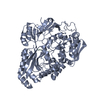
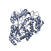
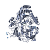
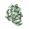
- PDB-6pu3: ABC transporter-associated periplasmic binding protein DppA from ... -
+ Open data

- Basic information
| Entry | Database: PDB / ID: 6pu3 | ||||||
|---|---|---|---|---|---|---|---|
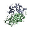
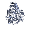
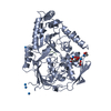
| Title | ABC transporter-associated periplasmic binding protein DppA from Helicobacter pylori | ||||||
 Components Components |
| ||||||
 Keywords Keywords | TRANSPORT PROTEIN / DppA / type II / periplasmic binding protein / Periplasmic peptide binding protein | ||||||
| Function / homology | Peptide/nickel binding protein, MppA-type / Solute-binding protein family 5 domain / Solute-binding protein family 5 / Bacterial extracellular solute-binding proteins, family 5 Middle / ATP-binding cassette (ABC) transporter complex / transmembrane transport / Heme-binding protein A Function and homology information Function and homology information | ||||||
| Biological species |   Helicobacter pylori (bacteria) Helicobacter pylori (bacteria) | ||||||
| Method |  X-RAY DIFFRACTION / SYNCHROTRON / MOLECULAR REPLACEMENT / Resolution: 1.8 Å X-RAY DIFFRACTION / SYNCHROTRON / MOLECULAR REPLACEMENT / Resolution: 1.8 Å | ||||||
 Authors Authors | Rahman, M.M. / Machuca, M.A. / Khan, M.F. / Barlow, C.K. / Schittenhelm, R.B. / Roujeinikova, A. | ||||||
 Citation Citation | Journal: J.Bacteriol. / Year: 2019 Title: Molecular Basis of Unexpected Specificity of ABC Transporter-Associated Substrate-Binding Protein DppA from Helicobacter pylori. Authors: Rahman, M.M. / Machuca, M.A. / Khan, M.F. / Barlow, C.K. / Schittenhelm, R.B. / Roujeinikova, A. | ||||||
| History |
|
- Structure visualization
| Structure viewer | Molecule: MolmilJmol/JSmol |
|---|
- Downloads & links
-Download
| PDBx/mmCIF format | 6pu3.cif.gz | 136.6 KB | Display | PDBx/mmCIF format |
|---|---|---|---|---|
| PDB format | pdb6pu3.ent.gz | 101.8 KB | Display | PDB format |
| PDBx/mmJSON format | 6pu3.json.gz | Tree view | PDBx/mmJSON format | |
| Others |  Other downloads Other downloads |
-Validation report
| Arichive directory | https://data.pdbj.org/pub/pdb/validation_reports/pu/6pu3ftp://data.pdbj.org/pub/pdb/validation_reports/pu/6pu3 | HTTPS FTP |
|---|
-Related structure data
| Related structure data |  6ofqC 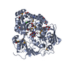 5f1qS C: citing same article ( S: Starting model for refinement |
|---|---|
| Similar structure data |
-Links
 PDBj
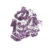
PDBj- Assembly
| Deposited unit | 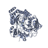
| ||||||||
|---|---|---|---|---|---|---|---|---|---|
| 1 | 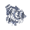
| ||||||||
| 2 |
| ||||||||
| Unit cell |
| ||||||||
| Details | The author determined assembly is a monomer. The tetrapeptide, Chain B, has co-purified with the protein and is not a part of the biological assembly |
-Components
| #1: Protein | Mass: 60510.992 Da / Num. of mol.: 1 Source method: isolated from a genetically manipulated source Source: (gene. exp.) Helicobacter pylori (bacteria) / Gene: hbpA, HPPMSS1_c01141 / Production host: |
|---|---|
| #2: Protein/peptide | Mass: 364.352 Da / Num. of mol.: 1 Source method: isolated from a genetically manipulated source Details: Probably co-purified with the protein / Source: (gene. exp.) |
| #3: Water | ChemComp-HOH /  Mass: 18.015 Da / Num. of mol.: 751 / Source method: isolated from a natural source / Formula: H2O Mass: 18.015 Da / Num. of mol.: 751 / Source method: isolated from a natural source / Formula: H2O |
-Experimental details
-Experiment
| Experiment | Method: X-RAY DIFFRACTION / Number of used crystals: 1 |
|---|
- Sample preparation
| Crystal | Density Matthews: 2.7 Å3/Da / Density % sol: 54.41 % |
|---|---|
| Crystal grow | Temperature: 293 K / Method: vapor diffusion, hanging drop Details: 23% w/v polyethylene glycol (PEG) 3350, 0.25 M tri-ammonium citrate |
-Data collection
| Diffraction | Mean temperature: 100.15 K / Serial crystal experiment: N |
|---|---|
| Diffraction source | Source: SYNCHROTRON / Site: Australian Synchrotron  / Beamline: MX1 / Wavelength: 0.9537 Å / Beamline: MX1 / Wavelength: 0.9537 Å |
| Detector | Type: ADSC QUANTUM 210r / Detector: CCD / Date: Jun 15, 2018 |
| Radiation | Protocol: SINGLE WAVELENGTH / Monochromatic (M) / Laue (L): M / Scattering type: x-ray |
| Radiation wavelength | Wavelength: 0.9537 Å / Relative weight: 1 |
| Reflection | Resolution: 1.8→46.12 Å / Num. obs: 59276 / % possible obs: 99 % / Redundancy: 3.6 % / CC1/2: 0.99 / Rmerge(I) obs: 0.102 / Net I/σ(I): 7.5 |
| Reflection shell | Resolution: 1.8→1.84 Å / Rmerge(I) obs: 0.348 / Num. unique obs: 3464 / CC1/2: 0.86 |
- Processing
| Software |
| ||||||||||||||||||||||||||||||||||||||||||||||||||||||||||||
|---|---|---|---|---|---|---|---|---|---|---|---|---|---|---|---|---|---|---|---|---|---|---|---|---|---|---|---|---|---|---|---|---|---|---|---|---|---|---|---|---|---|---|---|---|---|---|---|---|---|---|---|---|---|---|---|---|---|---|---|---|---|
| Refinement | Method to determine structure: MOLECULAR REPLACEMENT Starting model: 5F1Q Resolution: 1.8→46.12 Å / Cor.coef. Fo:Fc: 0.963 / Cor.coef. Fo:Fc free: 0.944 / SU B: 2.21 / SU ML: 0.067 / Cross valid method: THROUGHOUT / σ(F): 0 / ESU R: 0.098 / ESU R Free: 0.098 Details: HYDROGENS HAVE BEEN ADDED IN THE RIDING POSITIONS U VALUES : REFINED INDIVIDUALLY
| ||||||||||||||||||||||||||||||||||||||||||||||||||||||||||||
| Solvent computation | Ion probe radii: 0.8 Å / Shrinkage radii: 0.8 Å / VDW probe radii: 1.2 Å | ||||||||||||||||||||||||||||||||||||||||||||||||||||||||||||
| Displacement parameters | Biso max: 85.22 Å2 / Biso mean: 13.636 Å2 / Biso min: 3.71 Å2
| ||||||||||||||||||||||||||||||||||||||||||||||||||||||||||||
| Refinement step | Cycle: final / Resolution: 1.8→46.12 Å
| ||||||||||||||||||||||||||||||||||||||||||||||||||||||||||||
| Refine LS restraints |
| ||||||||||||||||||||||||||||||||||||||||||||||||||||||||||||
| LS refinement shell | Resolution: 1.8→1.847 Å / Rfactor Rfree error: 0 / Total num. of bins used: 20
|
