 Movie
Movie Controller

[English] 日本語
 Yorodumi
- PDB-2zl6: Atomic resolution structural characterization of recognition of h... -
+ Open data

- Basic information
| Entry | Database: PDB / ID: 2zl6 | |||||||||
|---|---|---|---|---|---|---|---|---|---|---|
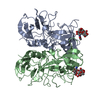
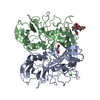
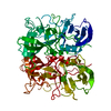
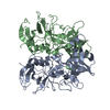
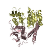
| Title | Atomic resolution structural characterization of recognition of histo-blood group antigens by Norwalk virus | |||||||||
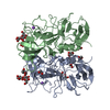
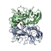
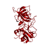
 Components Components | 58 kd capsid protein | |||||||||
 Keywords Keywords | VIRAL PROTEIN / Norovirus / Norwalk virus / HBGA / histo-blood group antigen / carbohydrate / VP1 / P-domain | |||||||||
| Function / homology |  Function and homology information Function and homology informationT=3 icosahedral viral capsid / host cell cytoplasm / identical protein binding Similarity search - Function | |||||||||
| Biological species |  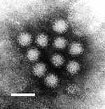 Norwalk virus Norwalk virus | |||||||||
| Method |  X-RAY DIFFRACTION / SYNCHROTRON / MOLECULAR REPLACEMENT / Resolution: 1.43 Å X-RAY DIFFRACTION / SYNCHROTRON / MOLECULAR REPLACEMENT / Resolution: 1.43 Å | |||||||||
 Authors Authors | Choi, J.M. / Huston, A.M. / Estes, M.K. / Prasad, B.V.V. | |||||||||
 Citation Citation | Journal: Proc.Natl.Acad.Sci.Usa / Year: 2008 Title: Atomic resolution structural characterization of recognition of histo-blood group antigens by Norwalk virus Authors: Choi, J.M. / Hutson, A.M. / Estes, M.K. / Prasad, B.V.V. | |||||||||
| History |
|
- Structure visualization
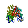
| Structure viewer | Molecule: MolmilJmol/JSmol |
|---|
- Downloads & links
-Download
| PDBx/mmCIF format | 2zl6.cif.gz | 142.3 KB | Display | PDBx/mmCIF format |
|---|---|---|---|---|
| PDB format | pdb2zl6.ent.gz | 108.6 KB | Display | PDB format |
| PDBx/mmJSON format | 2zl6.json.gz | Tree view | PDBx/mmJSON format | |
| Others |  Other downloads Other downloads |
-Validation report
| Arichive directory | https://data.pdbj.org/pub/pdb/validation_reports/zl/2zl6ftp://data.pdbj.org/pub/pdb/validation_reports/zl/2zl6 | HTTPS FTP |
|---|
-Related structure data
| Related structure data |  2zl5C  2zl7C 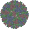 1ihmS C: citing same article ( S: Starting model for refinement |
|---|---|
| Similar structure data |

-Links
 PDBj
PDBj

- Assembly
| Deposited unit | 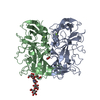
| |||||||||
|---|---|---|---|---|---|---|---|---|---|---|
| 1 |
| |||||||||
| Unit cell |
| |||||||||
| Components on special symmetry positions |
|
-Components
| #1: Protein | Mass: 31529.301 Da / Num. of mol.: 2 / Fragment: P-domain Source method: isolated from a genetically manipulated source Source: (gene. exp.) Norwalk virus / Strain: G.I / Gene: ORF2 / Plasmid: pHisMal-C2ET / Production host:  #2: Polysaccharide | alpha-L-fucopyranose-(1-2)-beta-D-galactopyranose-(1-3)-2-acetamido-2-deoxy-beta-D-glucopyranose-(1- ...alpha-L-fucopyranose-(1-2)-beta-D-galactopyranose-(1-3)-2-acetamido-2-deoxy-beta-D-glucopyranose-(1-3)-beta-D-galactopyranose-(1-4)-beta-D-glucopyranose | Source method: isolated from a genetically manipulated source #3: Chemical |   Mass: 24.305 Da / Num. of mol.: 2 / Source method: obtained synthetically / Formula: Mg Mass: 24.305 Da / Num. of mol.: 2 / Source method: obtained synthetically / Formula: Mg#4: Chemical | ChemComp-ACT / |   Mass: 59.044 Da / Num. of mol.: 1 / Source method: obtained synthetically / Formula: C2H3O2 Mass: 59.044 Da / Num. of mol.: 1 / Source method: obtained synthetically / Formula: C2H3O2#5: Water | ChemComp-HOH / |  Mass: 18.015 Da / Num. of mol.: 823 / Source method: isolated from a natural source / Formula: H2O Mass: 18.015 Da / Num. of mol.: 823 / Source method: isolated from a natural source / Formula: H2O |
|---|
-Experimental details
-Experiment
| Experiment | Method: X-RAY DIFFRACTION / Number of used crystals: 1 |
|---|
- Sample preparation
| Crystal | Density Matthews: 2.6 Å3/Da / Density % sol: 52.71 % |
|---|---|
| Crystal grow | Temperature: 293 K / Method: vapor diffusion, hanging drop / pH: 3.8 Details: 0.1M Citric Acid(pH 3.8), 0.2M Ammonium Nitrate, 20% PEG 3350, VAPOR DIFFUSION, HANGING DROP, temperature 293K |
-Data collection
| Diffraction | Mean temperature: 100 K |
|---|---|
| Diffraction source | Source: SYNCHROTRON / Site: APS  / Beamline: 19-ID / Wavelength: 0.97942 Å / Beamline: 19-ID / Wavelength: 0.97942 Å |
| Detector | Type: ADSC QUANTUM 315 / Detector: CCD / Date: Aug 5, 2007 |
| Radiation | Monochromator: Rosenbaum-Rock high-resolution double-crystal monochromator Protocol: SINGLE WAVELENGTH / Monochromatic (M) / Laue (L): M / Scattering type: x-ray |
| Radiation wavelength | Wavelength: 0.97942 Å / Relative weight: 1 |
| Reflection | Resolution: 1.43→41.73 Å / Num. all: 121668 / Num. obs: 121303 / % possible obs: 99.7 % / Observed criterion σ(F): 2 / Observed criterion σ(I): 2 / Redundancy: 5.38 % / Rmerge(I) obs: 0.07 / Net I/σ(I): 8.9 |
| Reflection shell | Resolution: 1.43→1.48 Å / Redundancy: 5.4 % / Rmerge(I) obs: 0.532 / Mean I/σ(I) obs: 2.5 / % possible all: 99.1 |
- Processing
| Software |
| ||||||||||||||||||||||||||||||||||||||||||||||||||||||||||||||||||||||||||||||||||||||||||
|---|---|---|---|---|---|---|---|---|---|---|---|---|---|---|---|---|---|---|---|---|---|---|---|---|---|---|---|---|---|---|---|---|---|---|---|---|---|---|---|---|---|---|---|---|---|---|---|---|---|---|---|---|---|---|---|---|---|---|---|---|---|---|---|---|---|---|---|---|---|---|---|---|---|---|---|---|---|---|---|---|---|---|---|---|---|---|---|---|---|---|---|
| Refinement | Method to determine structure: MOLECULAR REPLACEMENT Starting model: PDB ENTRY 1ihm Resolution: 1.43→40.45 Å / Cor.coef. Fo:Fc: 0.967 / Cor.coef. Fo:Fc free: 0.96 / SU B: 1.075 / SU ML: 0.042 / Cross valid method: THROUGHOUT / σ(F): 0 / σ(I): 0 / ESU R: 0.064 / ESU R Free: 0.064 / Stereochemistry target values: MAXIMUM LIKELIHOOD / Details: HYDROGENS HAVE BEEN ADDED IN THE RIDING POSITIONS
| ||||||||||||||||||||||||||||||||||||||||||||||||||||||||||||||||||||||||||||||||||||||||||
| Solvent computation | Ion probe radii: 0.8 Å / Shrinkage radii: 0.8 Å / VDW probe radii: 1.2 Å / Solvent model: MASK | ||||||||||||||||||||||||||||||||||||||||||||||||||||||||||||||||||||||||||||||||||||||||||
| Displacement parameters | Biso mean: 18.633 Å2
| ||||||||||||||||||||||||||||||||||||||||||||||||||||||||||||||||||||||||||||||||||||||||||
| Refinement step | Cycle: LAST / Resolution: 1.43→40.45 Å
| ||||||||||||||||||||||||||||||||||||||||||||||||||||||||||||||||||||||||||||||||||||||||||
| Refine LS restraints |
| ||||||||||||||||||||||||||||||||||||||||||||||||||||||||||||||||||||||||||||||||||||||||||
| LS refinement shell | Resolution: 1.43→1.467 Å / Total num. of bins used: 20
|
