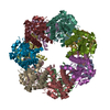
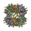
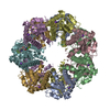
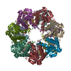
Mass: 21547.674 Da / Num. of mol.: 14 Source method: isolated from a genetically manipulated source Source: (gene. exp.) Helicobacter pylori (bacteria) / Gene: clpP / Plasmid: pET_22b(+) / Species (production host): Escherichia coli / Production host: Escherichia coli BL21(DE3) (bacteria) / Strain (production host): BL21(DE3) / References: UniProt: P56156, endopeptidase Clp
#2: Protein/peptide
Apeptidesubstrate-NVLGFTQ
Mass: 777.865 Da / Num. of mol.: 8 / Source method: obtained synthetically / Details: The peptide was chemically synthesized.
#3: Protein/peptide
Apeptidesubstrate-NVLGFTQforChainRandS
Mass: 613.749 Da / Num. of mol.: 2 / Source method: obtained synthetically / Details: The peptide was chemically synthesized.
Sequence details
BECAUSE THE ELECTRON DENSITIES FOR ALL OF SIDE CHAINS IN CHAIN R AND S WERE POOR, THE AUTHORS WERE ...BECAUSE THE ELECTRON DENSITIES FOR ALL OF SIDE CHAINS IN CHAIN R AND S WERE POOR, THE AUTHORS WERE UNABLE TO ASSIGN SIDE CHAINS OF THEM. IN ADDITION, RESIDUE NUMBERS ARE ARBITRARY. THE FOLLOWING IS THE ONE-LETTER SEQUENCE FOR THE PROTEIN IN CHAIN R AND S MODELED IN THIS STRUCTURE, NVLGFTQ.
-
Experimental details
-
Experiment
Experiment
Method: X-RAY DIFFRACTION / Number of used crystals: 1
-
Sample preparation
Crystal
Density Matthews: 2.34 Å3/Da / Density % sol: 47.45 %
In the structure databanks used in Yorodumi, some data are registered as the other names, "COVID-19 virus" and "2019-nCoV". Here are the details of the virus and the list of structure data.
Jan 31, 2019. EMDB accession codes are about to change! (news from PDBe EMDB page)
EMDB accession codes are about to change! (news from PDBe EMDB page)
The allocation of 4 digits for EMDB accession codes will soon come to an end. Whilst these codes will remain in use, new EMDB accession codes will include an additional digit and will expand incrementally as the available range of codes is exhausted. The current 4-digit format prefixed with “EMD-” (i.e. EMD-XXXX) will advance to a 5-digit format (i.e. EMD-XXXXX), and so on. It is currently estimated that the 4-digit codes will be depleted around Spring 2019, at which point the 5-digit format will come into force.
The EM Navigator/Yorodumi systems omit the EMD- prefix.
Related info.:Q: What is EMD? / ID/Accession-code notation in Yorodumi/EM Navigator
Yorodumi is a browser for structure data from EMDB, PDB, SASBDB, etc.
This page is also the successor to EM Navigator detail page, and also detail information page/front-end page for Omokage search.
The word "yorodu" (or yorozu) is an old Japanese word meaning "ten thousand". "mi" (miru) is to see.
Related info.:EMDB / PDB / SASBDB / Comparison of 3 databanks / Yorodumi Search / Aug 31, 2016. New EM Navigator & Yorodumi / Yorodumi Papers / Jmol/JSmol / Function and homology information / Changes in new EM Navigator and Yorodumi
 Movie
Movie Controller

 Yorodumi
 Open data
 Basic information
 Components
 Keywords
 Function and homology information
 Helicobacter pylori (bacteria)
 X-RAY DIFFRACTION /
  Authors
 Citation
 Structure visualization
 Downloads & links
 Other downloads
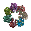


 PDBj
PDBj Assembly
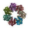
 Sample preparation
 / Beamline: AR-NW12A / Wavelength: 1 Å
 Processing