 Movie
Movie Controller

[English] 日本語
 Yorodumi
- PDB-2zho: Crystal structure of the regulatory subunit of aspartate kinase f... -
+ Open data

- Basic information
| Entry | Database: PDB / ID: 2zho | ||||||
|---|---|---|---|---|---|---|---|
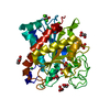
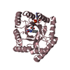
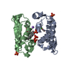
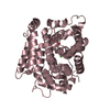
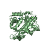
| Title | Crystal structure of the regulatory subunit of aspartate kinase from Thermus thermophilus (ligand free form) | ||||||
 Components Components | Aspartokinase | ||||||
 Keywords Keywords | TRANSFERASE / regulatory domain / ACT domain / Alternative initiation / Amino-acid biosynthesis / Diaminopimelate biosynthesis / Kinase / Lysine biosynthesis | ||||||
| Function / homology |  Function and homology information Function and homology informationaspartate kinase / aspartate kinase activity / L-homoserine biosynthetic process / L-threonine biosynthetic process / : / : / ATP binding / cytosol Similarity search - Function | ||||||
| Biological species |   Thermus thermophilus (bacteria) Thermus thermophilus (bacteria) | ||||||
| Method |  X-RAY DIFFRACTION / SYNCHROTRON / MOLECULAR REPLACEMENT / Resolution: 2.98 Å X-RAY DIFFRACTION / SYNCHROTRON / MOLECULAR REPLACEMENT / Resolution: 2.98 Å | ||||||
 Authors Authors | Yoshida, A. / Tomita, T. / Kuzuyama, T. / Nishiyama, M. | ||||||
 Citation Citation | Journal: Febs J. / Year: 2009 Title: Crystal structures of the regulatory subunit of Thr-sensitive aspartate kinase from Thermus thermophilus Authors: Yoshida, A. / Tomita, T. / Kono, H. / Fushinobu, S. / Kuzuyama, T. / Nishiyama, M. #1: Journal: J.Biosci.Bioeng. / Year: 1999 Title: Kinetic and mutation analyses of aspartate kinase from Thermus flavus Authors: Kobashi, N. / Nishiyama, M. / Tanokura, M. #2: Journal: Microbiology / Year: 1995 Title: An operon encoding aspartokinase and purine phosphoribosyltransferase in Thermus flavus Authors: Nishiyama, M. / Kukimoto, M. / Beppu, T. / Horinouchi, S. | ||||||
| History |
|
- Structure visualization
| Structure viewer | Molecule: MolmilJmol/JSmol |
|---|
- Downloads & links
-Download
| PDBx/mmCIF format | 2zho.cif.gz | 170.5 KB | Display | PDBx/mmCIF format |
|---|---|---|---|---|
| PDB format | pdb2zho.ent.gz | 136.4 KB | Display | PDB format |
| PDBx/mmJSON format | 2zho.json.gz | Tree view | PDBx/mmJSON format | |
| Others |  Other downloads Other downloads |
-Validation report
| Arichive directory | https://data.pdbj.org/pub/pdb/validation_reports/zh/2zhoftp://data.pdbj.org/pub/pdb/validation_reports/zh/2zho | HTTPS FTP |
|---|
-Related structure data
| Related structure data |  2dt9SC S: Starting model for refinement C: citing same article ( |
|---|---|
| Similar structure data |


-Links
 PDBj
PDBj

- Assembly
| Deposited unit | 
| ||||||||
|---|---|---|---|---|---|---|---|---|---|
| 1 | 
| ||||||||
| 2 | 
| ||||||||
| 3 | 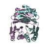
| ||||||||
| Unit cell |
|
-Components
| #1: Protein | Mass: 17738.367 Da / Num. of mol.: 6 Fragment: regulatory subunit, Aspartokinase subunit alpha and beta, UNP residues 245-405 Source method: isolated from a genetically manipulated source Source: (gene. exp.) Thermus thermophilus (bacteria) / Strain: AT-62 / Gene: ask / Plasmid: pET26b(+) / Production host: #2: Water | ChemComp-HOH / |  Mass: 18.015 Da / Num. of mol.: 79 / Source method: isolated from a natural source / Formula: H2O Mass: 18.015 Da / Num. of mol.: 79 / Source method: isolated from a natural source / Formula: H2O |
|---|
-Experimental details
-Experiment
| Experiment | Method: X-RAY DIFFRACTION / Number of used crystals: 1 |
|---|
- Sample preparation
| Crystal | Density Matthews: 2.72 Å3/Da / Density % sol: 54.74 % |
|---|---|
| Crystal grow | Temperature: 293 K / Method: vapor diffusion, hanging drop / pH: 5 Details: 1.6M sodium chloride, 0.1M Sodium citrate, pH 5.0, VAPOR DIFFUSION, HANGING DROP, temperature 293K |
-Data collection
| Diffraction | Mean temperature: 95 K |
|---|---|
| Diffraction source | Source: SYNCHROTRON / Site: Photon Factory  / Beamline: AR-NW12A / Wavelength: 1 Å / Beamline: AR-NW12A / Wavelength: 1 Å |
| Detector | Type: ADSC QUANTUM 210 / Detector: CCD / Date: Jun 1, 2006 / Details: mirror |
| Radiation | Monochromator: Si(111) double crystal monochromator / Protocol: SINGLE WAVELENGTH / Monochromatic (M) / Laue (L): M / Scattering type: x-ray |
| Radiation wavelength | Wavelength: 1 Å / Relative weight: 1 |
| Reflection | Resolution: 2.98→46.41 Å / Num. all: 22878 / Num. obs: 22878 / % possible obs: 99.8 % / Redundancy: 2.9 % / Biso Wilson estimate: 67.7 Å2 / Rmerge(I) obs: 0.092 / Rsym value: 0.092 / Net I/σ(I): 21 |
| Reflection shell | Resolution: 2.98→3.09 Å / Redundancy: 2.9 % / Mean I/σ(I) obs: 3.23 / Rsym value: 0.371 / % possible all: 100 |
- Processing
| Software |
| |||||||||||||||||||||||||
|---|---|---|---|---|---|---|---|---|---|---|---|---|---|---|---|---|---|---|---|---|---|---|---|---|---|---|
| Refinement | Method to determine structure: MOLECULAR REPLACEMENT Starting model: PDB ENTRY 2DT9 Resolution: 2.98→46.41 Å / Rfactor Rfree error: 0.008 / Data cutoff high absF: 2406887.39 / Data cutoff low absF: 0 / Isotropic thermal model: RESTRAINED / Cross valid method: THROUGHOUT / σ(F): 0 / Stereochemistry target values: Engh & Huber
| |||||||||||||||||||||||||
| Solvent computation | Solvent model: FLAT MODEL / Bsol: 36.2789 Å2 / ksol: 0.309011 e/Å3 | |||||||||||||||||||||||||
| Displacement parameters | Biso mean: 56.1 Å2
| |||||||||||||||||||||||||
| Refine analyze |
| |||||||||||||||||||||||||
| Refinement step | Cycle: LAST / Resolution: 2.98→46.41 Å
| |||||||||||||||||||||||||
| Refine LS restraints |
| |||||||||||||||||||||||||
| LS refinement shell | Resolution: 2.98→3.17 Å / Rfactor Rfree error: 0.025 / Total num. of bins used: 6
| |||||||||||||||||||||||||
| Xplor file |
|
