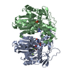
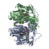
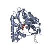
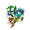
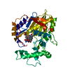
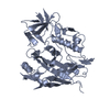
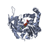
ジャーナル: EMBO J / 年: 2008 タイトル: Molecular structure of the ParM polymer and the mechanism leading to its nucleotide-driven dynamic instability. 著者: David Popp / Akihiro Narita / Toshiro Oda / Tetsuro Fujisawa / Hiroshi Matsuo / Yasushi Nitanai / Mitsusada Iwasa / Kayo Maeda / Hirofumi Onishi / Yuichiro Maéda / 要旨: ParM is a prokaryotic actin homologue, which ensures even plasmid segregation before bacterial cell division. In vivo, ParM forms a labile filament bundle that is reminiscent of the more complex ...ParM is a prokaryotic actin homologue, which ensures even plasmid segregation before bacterial cell division. In vivo, ParM forms a labile filament bundle that is reminiscent of the more complex spindle formed by microtubules partitioning chromosomes in eukaryotic cells. However, little is known about the underlying structural mechanism of DNA segregation by ParM filaments and the accompanying dynamic instability. Our biochemical, TIRF microscopy and high-pressure SAX observations indicate that polymerization and disintegration of ParM filaments is driven by GTP rather than ATP and that ParM acts as a GTP-driven molecular switch similar to a G protein. Image analysis of electron micrographs reveals that the ParM filament is a left-handed helix, opposed to the right-handed actin polymer. Nevertheless, the intersubunit contacts are similar to those of actin. Our atomic model of the ParM-GMPPNP filament, which also fits well to X-ray fibre diffraction patterns from oriented gels, can explain why after nucleotide release, large conformational changes of the protomer lead to a breakage of intra- and interstrand interactions, and thus to the observed disintegration of the ParM filament after DNA segregation.
濃度: 0.13 mg/ml / 包埋: NO / シャドウイング: NO / 染色: YES / 凍結: NO
染色
タイプ: NEGATIVE / 染色剤: Uranyl Acetate
試料支持
詳細: Carbon
-
電子顕微鏡撮影
顕微鏡
モデル: JEOL 2010HC / 日付: 2007年1月1日
電子銃
電子線源: LAB6 / 加速電圧: 100 kV / 照射モード: FLOOD BEAM
電子レンズ
モード: BRIGHT FIELD / 倍率(公称値): 40000 X / 最大 デフォーカス(公称値): 10300 nm / 最小 デフォーカス(公称値): 3700 nm
試料ホルダ
温度: 300 K / 傾斜角・最大: 0 ° / 傾斜角・最小: 0 °
撮影
電子線照射量: 12 e/Å2 / フィルム・検出器のモデル: GENERIC FILM
画像スキャン
デジタル画像の数: 2085
放射
プロトコル: SINGLE WAVELENGTH / 単色(M)・ラウエ(L): M
放射波長
相対比: 1
-
解析
EMソフトウェア
名称: EOS / カテゴリ: 3次元再構成
CTF補正
詳細: Phase and Amplitude
3次元再構成
手法: Single particle analysis / 解像度: 23 Å / 粒子像の数: 2085 / ピクセルサイズ(公称値): 4.011 Å / ピクセルサイズ(実測値): 4.011 Å 詳細: This data was achieved by negative staining experiments 対称性のタイプ: HELICAL
 ムービー
ムービー コントローラー


 データを開く
 基本情報
基本情報 要素
要素 キーワード
キーワード 機能・相同性情報
 データ登録者
 引用
引用
 構造の表示
構造の表示 ダウンロードとリンク
 その他のダウンロード
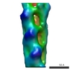


 PDBj
PDBj 集合体
集合体

 分子量: 24.305 Da / 分子数: 1 / 由来タイプ: 合成 / 式: Mg
 分子量: 427.201 Da / 分子数: 1 / 由来タイプ: 合成 / 式: C10H15N5O10P2 / コメント: ADP, エネルギー貯蔵分子*YM
 試料調製
試料調製 電子顕微鏡撮影
 解析
解析