 Movie
Movie Controller


+ Open data

- Basic information
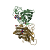
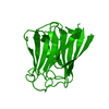
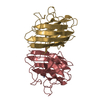
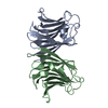
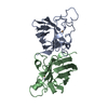
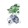
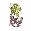
| Entry | Database: PDB / ID: 2zgp | ||||||
|---|---|---|---|---|---|---|---|
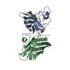
| Title | Crystal structure of Agrocybe aegerita lectin AAL mutant I25G | ||||||
 Components Components | Anti-tumor lectin | ||||||
 Keywords Keywords | HYDROLASE / galectin / jelly role / Apoptosis / Nuclease | ||||||
| Function / homology |  Function and homology information Function and homology informationDNA nuclease activity / Hydrolases; Acting on ester bonds; Endodeoxyribonucleases producing 5'-phosphomonoesters / polysaccharide binding / positive regulation of apoptotic process / hydrolase activity / apoptotic process Similarity search - Function | ||||||
| Biological species |  Agrocybe aegerita (fungus) Agrocybe aegerita (fungus) | ||||||
| Method |  X-RAY DIFFRACTION / MOLECULAR REPLACEMENT / Resolution: 2.7 Å X-RAY DIFFRACTION / MOLECULAR REPLACEMENT / Resolution: 2.7 Å | ||||||
 Authors Authors | Yang, N. / Li, D.F. / Wang, D.C. | ||||||
 Citation Citation | Journal: J.Mol.Biol. / Year: 2009 Title: Structural basis for the tumor cell apoptosis-inducing activity of an antitumor lectin from the edible mushroom Agrocybe aegerita Authors: Yang, N. / Li, D.F. / Feng, L. / Xiang, Y. / Liu, W. / Sun, H. / Wang, D.C. | ||||||
| History |
|
- Structure visualization
| Structure viewer | Molecule: MolmilJmol/JSmol |
|---|
- Downloads & links
-Download
| PDBx/mmCIF format | 2zgp.cif.gz | 72.4 KB | Display | PDBx/mmCIF format |
|---|---|---|---|---|
| PDB format | pdb2zgp.ent.gz | 53.5 KB | Display | PDB format |
| PDBx/mmJSON format | 2zgp.json.gz | Tree view | PDBx/mmJSON format | |
| Others |  Other downloads Other downloads |
-Validation report
| Arichive directory | https://data.pdbj.org/pub/pdb/validation_reports/zg/2zgpftp://data.pdbj.org/pub/pdb/validation_reports/zg/2zgp | HTTPS FTP |
|---|
-Related structure data
| Related structure data | 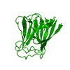 2zgkC 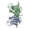 2zglSC  2zgmC 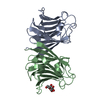 2zgnC 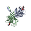 2zgoC 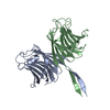 2zgqC  2zgrC  2zgsC  2zgtC  2zguC C: citing same article ( S: Starting model for refinement |
|---|---|
| Similar structure data |


-Links
 PDBj
PDBj- Assembly
| Deposited unit | 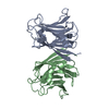
| ||||||||
|---|---|---|---|---|---|---|---|---|---|
| 1 |
| ||||||||
| Unit cell |
|
-Components
| #1: Protein | Mass: 18124.045 Da / Num. of mol.: 2 / Mutation: I25G Source method: isolated from a genetically manipulated source Source: (gene. exp.) Agrocybe aegerita (fungus) / Gene: AAL / Plasmid: pET22b / Production host:  References: UniProt: Q6WY08, Hydrolases; Acting on ester bonds; Endodeoxyribonucleases producing 5'-phosphomonoesters #2: Water | ChemComp-HOH / |  Mass: 18.015 Da / Num. of mol.: 78 / Source method: isolated from a natural source / Formula: H2O Mass: 18.015 Da / Num. of mol.: 78 / Source method: isolated from a natural source / Formula: H2OSequence details | THIS SEQUENCE IS ALLELE OF UNP Q6WY08. | |
|---|
-Experimental details
-Experiment
| Experiment | Method: X-RAY DIFFRACTION / Number of used crystals: 1 |
|---|
- Sample preparation
| Crystal | Density Matthews: 2.65 Å3/Da / Density % sol: 53.63 % |
|---|---|
| Crystal grow | Temperature: 298 K / Method: vapor diffusion, hanging drop / pH: 5.5 Details: 25% PEG3350, pH 5.5, VAPOR DIFFUSION, HANGING DROP, temperature 298K |
-Data collection
| Diffraction | Mean temperature: 98 K |
|---|---|
| Diffraction source | Source: ROTATING ANODE / Type: RIGAKU FR-E+ SUPERBRIGHT / Wavelength: 1.5418 Å |
| Detector | Type: RIGAKU RAXIS IV++ / Detector: IMAGE PLATE / Date: May 15, 2007 |
| Radiation | Protocol: SINGLE WAVELENGTH / Monochromatic (M) / Laue (L): M / Scattering type: x-ray |
| Radiation wavelength | Wavelength: 1.5418 Å / Relative weight: 1 |
| Reflection | Resolution: 2.7→31.98 Å / Num. all: 10538 / Num. obs: 10538 / % possible obs: 99.9 % / Observed criterion σ(F): 0 / Observed criterion σ(I): 0 / Redundancy: 3.6 % / Biso Wilson estimate: 41.9 Å2 / Rmerge(I) obs: 0.114 / Rsym value: 0.071 / Net I/σ(I): 12.6 |
| Reflection shell | Resolution: 2.7→2.85 Å / Redundancy: 3.6 % / Rmerge(I) obs: 0.322 / Mean I/σ(I) obs: 3.8 / Num. unique all: 1546 / Rsym value: 0.197 / % possible all: 100 |
- Processing
| Software |
| ||||||||||||||||||||||||||||||||||||||||||||||||||||||||||||
|---|---|---|---|---|---|---|---|---|---|---|---|---|---|---|---|---|---|---|---|---|---|---|---|---|---|---|---|---|---|---|---|---|---|---|---|---|---|---|---|---|---|---|---|---|---|---|---|---|---|---|---|---|---|---|---|---|---|---|---|---|---|
| Refinement | Method to determine structure: MOLECULAR REPLACEMENT Starting model: 2ZGL Resolution: 2.7→31.98 Å / Rfactor Rfree error: 0.008 / Data cutoff high absF: 780838.11 / Data cutoff low absF: 0 / Isotropic thermal model: RESTRAINED / Cross valid method: THROUGHOUT / σ(F): 2 / σ(I): 2 / Stereochemistry target values: Engh & Huber / Details: BULK SOLVENT MODEL USED
| ||||||||||||||||||||||||||||||||||||||||||||||||||||||||||||
| Solvent computation | Solvent model: FLAT MODEL / Bsol: 20.3066 Å2 / ksol: 0.35 e/Å3 | ||||||||||||||||||||||||||||||||||||||||||||||||||||||||||||
| Displacement parameters | Biso mean: 16.7 Å2
| ||||||||||||||||||||||||||||||||||||||||||||||||||||||||||||
| Refine analyze |
| ||||||||||||||||||||||||||||||||||||||||||||||||||||||||||||
| Refinement step | Cycle: LAST / Resolution: 2.7→31.98 Å
| ||||||||||||||||||||||||||||||||||||||||||||||||||||||||||||
| Refine LS restraints |
| ||||||||||||||||||||||||||||||||||||||||||||||||||||||||||||
| LS refinement shell | Resolution: 2.7→2.87 Å / Rfactor Rfree error: 0.03 / Total num. of bins used: 6
| ||||||||||||||||||||||||||||||||||||||||||||||||||||||||||||
| Xplor file |
|
