 Movie
Movie Controller

[English] 日本語
 Yorodumi
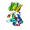
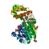
- PDB-2zcu: Crystal structure of a new type of NADPH-dependent quinone oxidor... -
+ Open data

- Basic information
| Entry | Database: PDB / ID: 2zcu | ||||||
|---|---|---|---|---|---|---|---|
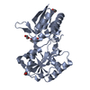
| Title | Crystal structure of a new type of NADPH-dependent quinone oxidoreductase (QOR2) from escherichia coli | ||||||
 Components Components | Uncharacterized oxidoreductase ytfG | ||||||
 Keywords Keywords | OXIDOREDUCTASE / ALPHA-BETA SANDWICH | ||||||
| Function / homology |  Function and homology information Function and homology informationNAD(P)H dehydrogenase (quinone) / NADPH dehydrogenase (quinone) activity / NADH dehydrogenase (quinone) (non-electrogenic) activity / oxidoreductase activity, acting on NAD(P)H, quinone or similar compound as acceptor / cytosol Similarity search - Function | ||||||
| Biological species |  | ||||||
| Method |  X-RAY DIFFRACTION / SYNCHROTRON / MAD / Resolution: 1.8 Å X-RAY DIFFRACTION / SYNCHROTRON / MAD / Resolution: 1.8 Å | ||||||
 Authors Authors | Kim, I.K. / Yim, H.S. / Kim, M.K. / Kim, D.W. / Kim, Y.M. / Cha, S.S. / Kang, S.O. | ||||||
 Citation Citation | Journal: J.Mol.Biol. / Year: 2008 Title: Crystal structure of a new type of NADPH-dependent quinone oxidoreductase (QOR2) from Escherichia coli Authors: Kim, I.K. / Yim, H.S. / Kim, M.K. / Kim, D.W. / Kim, Y.M. / Cha, S.S. / Kang, S.O. | ||||||
| History |
|
- Structure visualization
| Structure viewer | Molecule: MolmilJmol/JSmol |
|---|
- Downloads & links
-Download
| PDBx/mmCIF format | 2zcu.cif.gz | 71.5 KB | Display | PDBx/mmCIF format |
|---|---|---|---|---|
| PDB format | pdb2zcu.ent.gz | 51.9 KB | Display | PDB format |
| PDBx/mmJSON format | 2zcu.json.gz | Tree view | PDBx/mmJSON format | |
| Others |  Other downloads Other downloads |
-Validation report
| Summary document | 2zcu_validation.pdf.gz | 425.9 KB | Display | wwPDB validaton report |
|---|---|---|---|---|
| Full document | 2zcu_full_validation.pdf.gz | 431.8 KB | Display | |
| Data in XML | 2zcu_validation.xml.gz | 17.3 KB | Display | |
| Data in CIF | 2zcu_validation.cif.gz | 26.6 KB | Display | |
| Arichive directory | https://data.pdbj.org/pub/pdb/validation_reports/zc/2zcuftp://data.pdbj.org/pub/pdb/validation_reports/zc/2zcu | HTTPS FTP |
-Related structure data


-Links
 PDBj
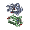
PDBj- Assembly
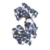
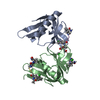
| Deposited unit | 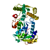
| ||||||||
|---|---|---|---|---|---|---|---|---|---|
| 1 |
| ||||||||
| 2 | 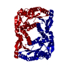
| ||||||||
| Unit cell |
|
-Components
| #1: Protein | Mass: 29762.406 Da / Num. of mol.: 1 Source method: isolated from a genetically manipulated source Source: (gene. exp.) |
|---|---|
| #2: Chemical | ChemComp-CU /   Mass: 63.546 Da / Num. of mol.: 1 / Source method: obtained synthetically / Formula: Cu Mass: 63.546 Da / Num. of mol.: 1 / Source method: obtained synthetically / Formula: Cu |
| #3: Water | ChemComp-HOH /  Mass: 18.015 Da / Num. of mol.: 419 / Source method: isolated from a natural source / Formula: H2O Mass: 18.015 Da / Num. of mol.: 419 / Source method: isolated from a natural source / Formula: H2O |
-Experimental details
-Experiment
| Experiment | Method: X-RAY DIFFRACTION / Number of used crystals: 1 |
|---|
- Sample preparation
| Crystal | Density Matthews: 2.39 Å3/Da / Density % sol: 48.8 % |
|---|---|
| Crystal grow | Temperature: 285 K / Method: evaporation / pH: 7.5 Details: PEG 4000, COPPER CHLRORIDE, AMMONIUM SULFATE, HEPES, pH 7.50, EVAPORATION, temperature 285K |
-Data collection
| Diffraction | Mean temperature: 100 K | |||||||||||||||
|---|---|---|---|---|---|---|---|---|---|---|---|---|---|---|---|---|
| Diffraction source | Source: SYNCHROTRON / Site: PAL/PLS  / Beamline: 6B / Wavelength: 0.97934, 0.97947, 0.97167, 1.12714 / Beamline: 6B / Wavelength: 0.97934, 0.97947, 0.97167, 1.12714 | |||||||||||||||
| Detector | Type: BRUKER PROTEUM 300 / Detector: CCD / Date: May 10, 2003 | |||||||||||||||
| Radiation | Protocol: MAD / Monochromatic (M) / Laue (L): M / Scattering type: x-ray | |||||||||||||||
| Radiation wavelength |
| |||||||||||||||
| Reflection | Resolution: 1.8→20 Å / Num. obs: 28204 / % possible obs: 97.9 % / Observed criterion σ(I): 0 / Biso Wilson estimate: 14.7 Å2 / Rsym value: 0.062 | |||||||||||||||
| Reflection shell | Resolution: 1.8→1.9 Å / Rsym value: 0.294 / % possible all: 92 |
- Processing
| Software |
| ||||||||||||||||||||||||||||||||||||||||||||||||||||||||||||
|---|---|---|---|---|---|---|---|---|---|---|---|---|---|---|---|---|---|---|---|---|---|---|---|---|---|---|---|---|---|---|---|---|---|---|---|---|---|---|---|---|---|---|---|---|---|---|---|---|---|---|---|---|---|---|---|---|---|---|---|---|---|
| Refinement | Method to determine structure: MAD / Resolution: 1.8→19.74 Å / Rfactor Rfree error: 0.005 / Data cutoff high absF: 435259.26 / Data cutoff low absF: 0 / Isotropic thermal model: RESTRAINED / Cross valid method: THROUGHOUT / σ(F): 0 / Stereochemistry target values: ENGH & HUBER
| ||||||||||||||||||||||||||||||||||||||||||||||||||||||||||||
| Solvent computation | Solvent model: FLAT MODEL / Bsol: 98.99 Å2 / ksol: 0.35 e/Å3 | ||||||||||||||||||||||||||||||||||||||||||||||||||||||||||||
| Displacement parameters | Biso mean: 23.8 Å2
| ||||||||||||||||||||||||||||||||||||||||||||||||||||||||||||
| Refine analyze |
| ||||||||||||||||||||||||||||||||||||||||||||||||||||||||||||
| Refinement step | Cycle: LAST / Resolution: 1.8→19.74 Å
| ||||||||||||||||||||||||||||||||||||||||||||||||||||||||||||
| Refine LS restraints |
| ||||||||||||||||||||||||||||||||||||||||||||||||||||||||||||
| LS refinement shell | Resolution: 1.8→1.91 Å / Rfactor Rfree error: 0.014 / Total num. of bins used: 6
| ||||||||||||||||||||||||||||||||||||||||||||||||||||||||||||
| Xplor file |
|
