Resolution: 1.8→29.934 Å / Num. obs: 54677 / % possible obs: 100 % / Redundancy: 7.3 % / Biso Wilson estimate: 17.486 Å2 / Rmerge(I) obs: 0.114 / Rsym value: 0.114 / Net I/σ(I): 5
Reflection shell
Resolution (Å)
Redundancy (%)
Rmerge(I) obs
Mean I/σ(I) obs
Num. measured all
Num. unique all
Rsym value
% possible all
1.8-1.85
7.3
0.558
1.4
29118
3969
0.558
100
1.85-1.9
7.3
0.453
1.7
28489
3880
0.453
100
1.9-1.95
7.3
0.374
2
27786
3784
0.374
100
1.95-2.01
7.4
0.301
2.5
26985
3662
0.301
100
2.01-2.08
7.3
0.252
3
26239
3570
0.252
100
2.08-2.15
7.4
0.214
3.5
25472
3449
0.214
100
2.15-2.23
7.4
0.188
4
24460
3321
0.188
100
2.23-2.32
7.4
0.173
4.2
23945
3257
0.173
100
2.32-2.43
7.4
0.154
4.8
22676
3077
0.154
100
2.43-2.55
7.4
0.132
5.4
21686
2944
0.132
100
2.55-2.68
7.3
0.119
6
20779
2829
0.119
100
2.68-2.85
7.3
0.107
6.3
19638
2677
0.107
100
2.85-3.04
7.3
0.096
6.9
18337
2507
0.096
100
3.04-3.29
7.3
0.085
7.6
17260
2380
0.085
100
3.29-3.6
7.2
0.073
8.6
15718
2170
0.073
100
3.6-4.02
7.2
0.068
9.1
14337
1991
0.068
100
4.02-4.65
7.1
0.067
8.8
12617
1780
0.067
100
4.65-5.69
7
0.071
8.4
10510
1511
0.071
100
5.69-8.05
6.7
0.079
7.4
8078
1209
0.079
100
8.05-29.93
6
0.07
7.2
4282
710
0.07
98
-
Phasing
Phasing
Method: MAD
-
Processing
Software
Name
Version
Classification
NB
REFMAC
5.4.0067
refinement
PHENIX
refinement
SHELX
phasing
MolProbity
3beta29
modelbuilding
SCALA
datascaling
PDB_EXTRACT
3.004
dataextraction
MOSFLM
datareduction
SHELXD
phasing
autoSHARP
phasing
Refinement
Method to determine structure: MAD / Resolution: 1.8→29.934 Å / Cor.coef. Fo:Fc: 0.958 / Cor.coef. Fo:Fc free: 0.943 / SU B: 2.34 / SU ML: 0.075 / Cross valid method: THROUGHOUT / σ(F): 0 / ESU R: 0.124 / ESU R Free: 0.12 Stereochemistry target values: MAXIMUM LIKELIHOOD WITH PHASES Details: 1. HYDROGENS HAVE BEEN ADDED IN THE RIDING POSITIONS. 2. CITRATE (CIT) AND PEG-4000 (1PE) MOLECULES FROM THE CRYSTALLIZATION/CRYO SOLUTIONS ARE MODELED.
Rfactor
Num. reflection
% reflection
Selection details
Rfree
0.208
2772
5.1 %
RANDOM
Rwork
0.168
-
-
-
obs
0.17
54610
99.96 %
-
Solvent computation
Ion probe radii: 0.8 Å / Shrinkage radii: 0.8 Å / VDW probe radii: 1.2 Å / Solvent model: BABINET MODEL WITH MASK
Displacement parameters
Biso mean: 16.944 Å2
Baniso -1
Baniso -2
Baniso -3
1-
0.36 Å2
0 Å2
0 Å2
2-
-
0.36 Å2
0 Å2
3-
-
-
-0.72 Å2
Refinement step
Cycle: LAST / Resolution: 1.8→29.934 Å
Protein
Nucleic acid
Ligand
Solvent
Total
Num. atoms
4254
0
29
680
4963
Refine LS restraints
Refine-ID
Type
Dev ideal
Dev ideal target
Number
X-RAY DIFFRACTION
r_bond_refined_d
0.016
0.022
4566
X-RAY DIFFRACTION
r_bond_other_d
0.002
0.02
3093
X-RAY DIFFRACTION
r_angle_refined_deg
1.739
1.965
6231
X-RAY DIFFRACTION
r_angle_other_deg
1.519
3
7581
X-RAY DIFFRACTION
r_dihedral_angle_1_deg
4.605
5
591
X-RAY DIFFRACTION
r_dihedral_angle_2_deg
32.396
24.36
211
X-RAY DIFFRACTION
r_dihedral_angle_3_deg
11.437
15
779
X-RAY DIFFRACTION
r_dihedral_angle_4_deg
14.327
15
24
X-RAY DIFFRACTION
r_chiral_restr
0.101
0.2
687
X-RAY DIFFRACTION
r_gen_planes_refined
0.01
0.021
5136
X-RAY DIFFRACTION
r_gen_planes_other
0.004
0.02
938
X-RAY DIFFRACTION
r_mcbond_it
1.375
2
2784
X-RAY DIFFRACTION
r_mcbond_other
0.296
2
1117
X-RAY DIFFRACTION
r_mcangle_it
2.439
4
4535
X-RAY DIFFRACTION
r_scbond_it
4.089
6
1782
X-RAY DIFFRACTION
r_scangle_it
5.898
8
1671
LS refinement shell
Resolution: 1.8→1.847 Å / Total num. of bins used: 20
Rfactor
Num. reflection
% reflection
Rfree
0.256
167
-
Rwork
0.192
3800
-
all
-
3967
-
obs
-
-
100 %
+
About Yorodumi
-
News
-
Feb 9, 2022. New format data for meta-information of EMDB entries
New format data for meta-information of EMDB entries
Version 3 of the EMDB header file is now the official format.
The previous official version 1.9 will be removed from the archive.
In the structure databanks used in Yorodumi, some data are registered as the other names, "COVID-19 virus" and "2019-nCoV". Here are the details of the virus and the list of structure data.
Jan 31, 2019. EMDB accession codes are about to change! (news from PDBe EMDB page)
EMDB accession codes are about to change! (news from PDBe EMDB page)
The allocation of 4 digits for EMDB accession codes will soon come to an end. Whilst these codes will remain in use, new EMDB accession codes will include an additional digit and will expand incrementally as the available range of codes is exhausted. The current 4-digit format prefixed with “EMD-” (i.e. EMD-XXXX) will advance to a 5-digit format (i.e. EMD-XXXXX), and so on. It is currently estimated that the 4-digit codes will be depleted around Spring 2019, at which point the 5-digit format will come into force.
The EM Navigator/Yorodumi systems omit the EMD- prefix.
Related info.:Q: What is EMD? / ID/Accession-code notation in Yorodumi/EM Navigator
Yorodumi is a browser for structure data from EMDB, PDB, SASBDB, etc.
This page is also the successor to EM Navigator detail page, and also detail information page/front-end page for Omokage search.
The word "yorodu" (or yorozu) is an old Japanese word meaning "ten thousand". "mi" (miru) is to see.
Related info.:EMDB / PDB / SASBDB / Comparison of 3 databanks / Yorodumi Search / Aug 31, 2016. New EM Navigator & Yorodumi / Yorodumi Papers / Jmol/JSmol / Function and homology information / Changes in new EM Navigator and Yorodumi
 Movie
Movie Controller

 Yorodumi
 Open data
 Basic information
 Components
 Keywords
 Function and homology information
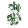
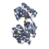
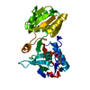
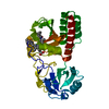
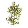
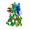
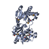
 Eubacterium rectale (bacteria)
 X-RAY DIFFRACTION /
  Authors
 Citation
 Structure visualization
 Downloads & links
 Other downloads
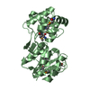


 PDBj
PDBj

 Assembly


 Mass: 238.278 Da / Num. of mol.: 1 / Source method: obtained synthetically / Formula: C10H22O6 / Comment: precipitant*YM
 Mass: 192.124 Da / Num. of mol.: 1 / Source method: obtained synthetically / Formula: C6H8O7
 Mass: 18.015 Da / Num. of mol.: 680 / Source method: isolated from a natural source / Formula: H2O
 Sample preparation
 / Beamline: BL9-2 / Wavelength: 0.97915,0.97929,0.91162
 Processing