 Movie
Movie Controller

[English] 日本語
 Yorodumi
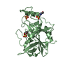
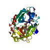
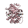
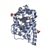
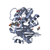
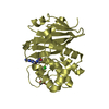
- PDB-2z8p: Structural basis for the catalytic mechanism of phosphothreonine lyase -
+ Open data

- Basic information
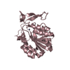
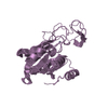
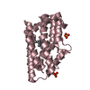
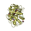
| Entry | Database: PDB / ID: 2z8p | ||||||
|---|---|---|---|---|---|---|---|
| Title | Structural basis for the catalytic mechanism of phosphothreonine lyase | ||||||
 Components Components |
| ||||||
 Keywords Keywords | LYASE / short three-helix bundle / distorted beta-strand sheet | ||||||
| Function / homology |  Function and homology information Function and homology informationLyases; Carbon-oxygen lyases; Acting on phosphates / lyase activity / extracellular region Similarity search - Function | ||||||
| Biological species |  Salmonella typhimurium (bacteria) Salmonella typhimurium (bacteria) | ||||||
| Method |  X-RAY DIFFRACTION / MOLECULAR REPLACEMENT / Resolution: 1.8 Å X-RAY DIFFRACTION / MOLECULAR REPLACEMENT / Resolution: 1.8 Å | ||||||
 Authors Authors | Chen, L. / Wang, H. / Gu, L. / Huang, N. / Zhou, J.M. / Chai, J. | ||||||
 Citation Citation | Journal: Nat.Struct.Mol.Biol. / Year: 2008 Title: Structural basis for the catalytic mechanism of phosphothreonine lyase. Authors: Chen, L. / Wang, H. / Zhang, J. / Gu, L. / Huang, N. / Zhou, J.M. / Chai, J. | ||||||
| History |
|
- Structure visualization
| Structure viewer | Molecule: MolmilJmol/JSmol |
|---|
- Downloads & links
-Download
| PDBx/mmCIF format | 2z8p.cif.gz | 65.5 KB | Display | PDBx/mmCIF format |
|---|---|---|---|---|
| PDB format | pdb2z8p.ent.gz | 46.8 KB | Display | PDB format |
| PDBx/mmJSON format | 2z8p.json.gz | Tree view | PDBx/mmJSON format | |
| Others |  Other downloads Other downloads |
-Validation report
| Summary document | 2z8p_validation.pdf.gz | 440.5 KB | Display | wwPDB validaton report |
|---|---|---|---|---|
| Full document | 2z8p_full_validation.pdf.gz | 446.7 KB | Display | |
| Data in XML | 2z8p_validation.xml.gz | 15 KB | Display | |
| Data in CIF | 2z8p_validation.cif.gz | 21.5 KB | Display | |
| Arichive directory | https://data.pdbj.org/pub/pdb/validation_reports/z8/2z8pftp://data.pdbj.org/pub/pdb/validation_reports/z8/2z8p | HTTPS FTP |
-Related structure data
| Related structure data | 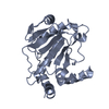 2z8mC 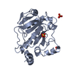 2z8nC  2z8oSC C: citing same article ( S: Starting model for refinement |
|---|---|
| Similar structure data |
-Links
 PDBj
PDBj- Assembly
| Deposited unit | 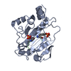
| ||||||||
|---|---|---|---|---|---|---|---|---|---|
| 1 |
| ||||||||
| Unit cell |
|
-Components
| #1: Protein | Mass: 27623.037 Da / Num. of mol.: 1 / Mutation: K136A Source method: isolated from a genetically manipulated source Source: (gene. exp.) Salmonella typhimurium (bacteria) / Plasmid: pGEX6p-1 / Species (production host): Escherichia coli / Production host: |
|---|---|
| #2: Protein/peptide | ( Mass: 869.704 Da / Num. of mol.: 1 / Source method: obtained synthetically / Details: The peptide was chemically synthesized. |
| #3: Water | ChemComp-HOH /  Mass: 18.015 Da / Num. of mol.: 259 / Source method: isolated from a natural source / Formula: H2O Mass: 18.015 Da / Num. of mol.: 259 / Source method: isolated from a natural source / Formula: H2O |
| Has protein modification | Y |
-Experimental details
-Experiment
| Experiment | Method: X-RAY DIFFRACTION / Number of used crystals: 1 |
|---|
- Sample preparation
| Crystal | Density Matthews: 2.31 Å3/Da / Density % sol: 46.84 % |
|---|---|
| Crystal grow | Temperature: 293 K / Method: vapor diffusion, hanging drop / pH: 7.5 Details: 17% PEG (MME) 2000, 100mM HEPES, pH 7.5, VAPOR DIFFUSION, HANGING DROP, temperature 293K |
-Data collection
| Diffraction | Mean temperature: 100 K |
|---|---|
| Diffraction source | Source: ROTATING ANODE / Type: RIGAKU RU300 / Wavelength: 1.5418 Å |
| Detector | Type: RIGAKU RAXIS IV / Detector: IMAGE PLATE / Date: Jan 22, 2007 |
| Radiation | Monochromator: Monochromator / Protocol: SINGLE WAVELENGTH / Monochromatic (M) / Laue (L): M / Scattering type: x-ray |
| Radiation wavelength | Wavelength: 1.5418 Å / Relative weight: 1 |
| Reflection | Resolution: 1.8→99 Å / Num. all: 25249 / Num. obs: 25173 / % possible obs: 99.7 % / Observed criterion σ(F): 0 / Observed criterion σ(I): 0 / Rmerge(I) obs: 0.052 / Net I/σ(I): 47.6 |
| Reflection shell | Resolution: 1.8→1.86 Å / Redundancy: 11.1 % / Rmerge(I) obs: 0.346 / Mean I/σ(I) obs: 8.5 / % possible all: 99.7 |
- Processing
| Software |
| |||||||||||||||||||||||||
|---|---|---|---|---|---|---|---|---|---|---|---|---|---|---|---|---|---|---|---|---|---|---|---|---|---|---|
| Refinement | Method to determine structure: MOLECULAR REPLACEMENT Starting model: 2Z8O Resolution: 1.8→20 Å / σ(F): 0 / σ(I): 0 / Stereochemistry target values: MAXIMUM LIKELIHOOD
| |||||||||||||||||||||||||
| Displacement parameters | Biso mean: 34.9473 Å2
| |||||||||||||||||||||||||
| Refinement step | Cycle: LAST / Resolution: 1.8→20 Å
| |||||||||||||||||||||||||
| Refine LS restraints |
| |||||||||||||||||||||||||
| LS refinement shell | Resolution: 1.8→1.82 Å
|
