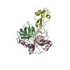
[English] 日本語
Yorodumi
- PDB-2z7f: Crystal structure of the complex of human neutrophil elastase wit... -

+
Open data


ID or keywords:

Loading...

-
Basic information

Entry
Database: PDB / ID: 2z7f
TitleCrystal structure of the complex of human neutrophil elastase with 1/2SLPI
Components
  • Antileukoproteinase
  • Leukocyte elastase
KeywordsHydrolase/Hydrolase inhibitor / serine protease / serine protease inhibitor / Disease mutation / Glycoprotein / Hydrolase / Zymogen / Secreted / Hydrolase-Hydrolase inhibitor COMPLEX
Function / homology
Function and homology information


modulation of process of another organism / leukocyte elastase / biosynthetic process of antibacterial peptides active against Gram-negative bacteria / neutrophil-mediated killing of fungus / negative regulation of chemotaxis / acute inflammatory response to antigenic stimulus / positive regulation of leukocyte tethering or rolling / response to yeast / leukocyte migration involved in inflammatory response / negative regulation of interleukin-8 production ...modulation of process of another organism / leukocyte elastase / biosynthetic process of antibacterial peptides active against Gram-negative bacteria / neutrophil-mediated killing of fungus / negative regulation of chemotaxis / acute inflammatory response to antigenic stimulus / positive regulation of leukocyte tethering or rolling / response to yeast / leukocyte migration involved in inflammatory response / negative regulation of interleukin-8 production / negative regulation of chemokine production / Antimicrobial peptides / endopeptidase inhibitor activity / negative regulation of viral genome replication / cytokine binding / pyroptotic inflammatory response / Activation of Matrix Metalloproteinases / neutrophil-mediated killing of gram-negative bacterium / Collagen degradation / extracellular matrix disassembly / Pyroptosis / phagocytosis / response to UV / transcription repressor complex / Degradation of the extracellular matrix / phagocytic vesicle / negative regulation of protein binding / secretory granule / Regulation of Complement cascade / positive regulation of interleukin-8 production / positive regulation of smooth muscle cell proliferation / positive regulation of MAP kinase activity / protein catabolic process / serine-type endopeptidase inhibitor activity / negative regulation of inflammatory response / intracellular calcium ion homeostasis / specific granule lumen / transcription corepressor activity / positive regulation of immune response / azurophil granule lumen / peptidase activity / heparin binding / antibacterial humoral response / collagen-containing extracellular matrix / protease binding / endopeptidase activity / response to lipopolysaccharide / defense response to bacterium / immune response / serine-type endopeptidase activity / innate immune response / mRNA binding / Neutrophil degranulation / Golgi apparatus / enzyme binding / negative regulation of transcription by RNA polymerase II / cell surface / proteolysis / DNA binding / extracellular space / extracellular exosome / extracellular region / cytoplasm / cytosol
Similarity search - Function
Elafin-like / R-elafin / WAP-type 'four-disulfide core' domain / Elafin-like superfamily / WAP-type (Whey Acidic Protein) 'four-disulfide core' / WAP-type 'four-disulfide core' domain profile. / Four-disulfide core domains / Few Secondary Structures / Irregular / Serine proteases, trypsin family, histidine active site ...Elafin-like / R-elafin / WAP-type 'four-disulfide core' domain / Elafin-like superfamily / WAP-type (Whey Acidic Protein) 'four-disulfide core' / WAP-type 'four-disulfide core' domain profile. / Four-disulfide core domains / Few Secondary Structures / Irregular / Serine proteases, trypsin family, histidine active site / Serine proteases, trypsin family, serine active site / Peptidase S1A, chymotrypsin family / Serine proteases, trypsin family, histidine active site. / Serine proteases, trypsin domain profile. / Serine proteases, trypsin family, serine active site. / Trypsin-like serine protease / Serine proteases, trypsin domain / Trypsin / Trypsin-like serine proteases / Thrombin, subunit H / Peptidase S1, PA clan, chymotrypsin-like fold / Peptidase S1, PA clan / Beta Barrel / Mainly Beta
Similarity search - Domain/homology
Antileukoproteinase / Neutrophil elastase
Similarity search - Component
Biological speciesHomo sapiens (human)
MethodX-RAY DIFFRACTION / SYNCHROTRON / MOLECULAR REPLACEMENT / Resolution: 1.7 Å
AuthorsTakimoto-Kamimura, M. / Fukushima, K.
CitationJournal: J.SYNCHROTRON RADIAT. / Year: 2008
Title: Complex of human neutrophil elastase with 1/2SLPI
Authors: Koizumi, M. / Fujino, A. / Fukushima, K. / Kamimura, T. / Takimoto-Kamimura, M.
History
DepositionAug 20, 2007Deposition site: PDBJ / Processing site: PDBJ
Revision 1.0Aug 26, 2008Provider: repository / Type: Initial release
Revision 1.1Jul 13, 2011Group: Non-polymer description / Version format compliance
Revision 2.0Jul 29, 2020Group: Atomic model / Data collection ...Atomic model / Data collection / Derived calculations / Structure summary
Category: atom_site / chem_comp ...atom_site / chem_comp / entity / pdbx_branch_scheme / pdbx_chem_comp_identifier / pdbx_entity_branch / pdbx_entity_branch_descriptor / pdbx_entity_branch_link / pdbx_entity_branch_list / pdbx_entity_nonpoly / pdbx_nonpoly_scheme / pdbx_struct_assembly_gen / pdbx_struct_special_symmetry / struct_asym / struct_conn / struct_site / struct_site_gen
Item: _atom_site.auth_asym_id / _atom_site.auth_seq_id ..._atom_site.auth_asym_id / _atom_site.auth_seq_id / _atom_site.label_asym_id / _atom_site.label_entity_id / _chem_comp.name / _chem_comp.type / _pdbx_struct_assembly_gen.asym_id_list / _pdbx_struct_special_symmetry.label_asym_id / _struct_conn.pdbx_leaving_atom_flag / _struct_conn.pdbx_role / _struct_conn.ptnr1_auth_asym_id / _struct_conn.ptnr1_auth_seq_id / _struct_conn.ptnr1_label_asym_id / _struct_conn.ptnr2_auth_asym_id / _struct_conn.ptnr2_auth_seq_id / _struct_conn.ptnr2_label_asym_id
Description: Carbohydrate remediation / Provider: repository / Type: Remediation

-
Structure visualization

Structure viewerMolecule:
MolmilJmol/JSmol

Downloads & links

-
Assembly

Deposited unit
E: Leukocyte elastase
I: Antileukoproteinase
hetero molecules


Theoretical massNumber of molelcules
Total (without water)29,5764
Polymers28,8422
Non-polymers7352
Water4,486249
1


  • Idetical with deposited unit
  • defined by author&software
TypeNameSymmetry operationNumber
identity operation1_555x,y,z1
Buried area2610 Å2
ΔGint2 kcal/mol
Surface area12700 Å2
MethodPISA
Unit cell
Length a, b, c (Å)106.638, 106.638, 55.117
Angle α, β, γ (deg.)90.00, 90.00, 90.00
Int Tables number90
Space group name H-MP4212
Components on special symmetry positions
IDModelComponents
11E-361-

HOH

-
Components

#1: Protein Leukocyte elastase / Elastase-2 / Neutrophil elastase / PMN elastase / Bone marrow serine protease / Medullasin / Human ...Elastase-2 / Neutrophil elastase / PMN elastase / Bone marrow serine protease / Medullasin / Human leukocyte elastase / HLE


Mass: 23318.982 Da / Num. of mol.: 1 / Fragment: Peptidase S1 domain
Source method: isolated from a genetically manipulated source
Source: (gene. exp.) Homo sapiens (human) / Tissue: sputum / Production host: Escherichia coli (E. coli) / References: UniProt: P08246, leukocyte elastase
#2: Protein/peptide Antileukoproteinase / ALP / Secretory leukocyte protease inhibitor / HUSI-1 / Seminal proteinase inhibitor / BLPI / Mucus ...ALP / Secretory leukocyte protease inhibitor / HUSI-1 / Seminal proteinase inhibitor / BLPI / Mucus proteinase inhibitor / MPI / WAP four-disulfide core domain protein 4 / Protease inhibitor WAP4


Mass: 5522.736 Da / Num. of mol.: 1 / Fragment: WAP 2 domain
Source method: isolated from a genetically manipulated source
Source: (gene. exp.) Homo sapiens (human) / Production host: Escherichia coli (E. coli) / References: UniProt: P03973
#3: Polysaccharide alpha-L-fucopyranose-(1-6)-2-acetamido-2-deoxy-beta-D-glucopyranose


Type: oligosaccharide / Mass: 367.349 Da / Num. of mol.: 2
Source method: isolated from a genetically manipulated source
DescriptorTypeProgram
LFucpa1-6DGlcpNAcb1-Glycam Condensed SequenceGMML 1.0
WURCS=2.0/2,2,1/[a2122h-1b_1-5_2*NCC/3=O][a1221m-1a_1-5]/1-2/a6-b1WURCSPDB2Glycan 1.1.0
[]{[(4+1)][b-D-GlcpNAc]{[(6+1)][a-L-Fucp]{}}}LINUCSPDB-CARE
#4: Water ChemComp-HOH / water


Mass: 18.015 Da / Num. of mol.: 249 / Source method: isolated from a natural source / Formula: H2O

-
Experimental details

-
Experiment

ExperimentMethod: X-RAY DIFFRACTION / Number of used crystals: 1

-
Sample preparation

CrystalDensity Matthews: 2.72 Å3/Da / Density % sol: 54.72 %
Crystal growTemperature: 293 K / Method: vapor diffusion, sitting drop / pH: 4.5
Details: 0.1M Na-Acetate, 2.0M Na-Formate, pH 4.5, VAPOR DIFFUSION, SITTING DROP, temperature 293.0K

-
Data collection

DiffractionMean temperature: 100 K
Diffraction sourceSource: SYNCHROTRON / Site: SPring-8 / Beamline: BL41XU / Wavelength: 1 Å
DetectorType: ADSC QUANTUM 315 / Detector: CCD / Date: Jun 2, 2005
RadiationProtocol: SINGLE WAVELENGTH / Monochromatic (M) / Laue (L): M / Scattering type: x-ray
Radiation wavelengthWavelength: 1 Å / Relative weight: 1
ReflectionResolution: 1.7→38.32 Å / Num. obs: 35374 / % possible obs: 99.5 % / Observed criterion σ(F): 2 / Observed criterion σ(I): 2 / Redundancy: 11.84 % / Rmerge(I) obs: 0.064 / Net I/σ(I): 18.5
Reflection shellResolution: 1.7→1.744 Å / Redundancy: 11.96 % / Mean I/σ(I) obs: 7.9 / Num. unique all: 134 / % possible all: 99.61

-
Processing

Software
NameVersionClassification
REFMAC5.2.0005refinement
CrystalCleardata collection
CrystalCleardata reduction
CrystalCleardata scaling
MOLREP(CCP4 6.01 suite)phasing
RefinementMethod to determine structure: MOLECULAR REPLACEMENT / Resolution: 1.7→38.32 Å / Cor.coef. Fo:Fc: 0.949 / Cor.coef. Fo:Fc free: 0.933 / SU B: 1.761 / SU ML: 0.061 / Cross valid method: THROUGHOUT / ESU R: 0.108 / ESU R Free: 0.105 / Stereochemistry target values: MAXIMUM LIKELIHOOD / Details: HYDROGENS HAVE BEEN ADDED IN THE RIDING POSITIONS
RfactorNum. reflection% reflectionSelection details
Rfree0.23062 1770 5 %RANDOM
Rwork0.20285 ---
obs0.20422 33575 99.45 %-
all-35374 --
Solvent computationIon probe radii: 0.8 Å / Shrinkage radii: 0.8 Å / VDW probe radii: 1.2 Å / Solvent model: MASK
Displacement parametersBiso mean: 20.882 Å2
Baniso -1Baniso -2Baniso -3
1-0.02 Å20 Å20 Å2
2--0.02 Å20 Å2
3----0.03 Å2
Refinement stepCycle: LAST / Resolution: 1.7→38.32 Å
ProteinNucleic acidLigandSolventTotal
Num. atoms2010 0 48 249 2307
Refine LS restraints
Refine-IDTypeDev idealDev ideal targetNumber
X-RAY DIFFRACTIONr_bond_refined_d0.0110.0222106
X-RAY DIFFRACTIONr_bond_other_d
X-RAY DIFFRACTIONr_angle_refined_deg1.2681.982863
X-RAY DIFFRACTIONr_angle_other_deg
X-RAY DIFFRACTIONr_dihedral_angle_1_deg5.8915266
X-RAY DIFFRACTIONr_dihedral_angle_2_deg31.48122.44286
X-RAY DIFFRACTIONr_dihedral_angle_3_deg12.91315332
X-RAY DIFFRACTIONr_dihedral_angle_4_deg17.4951522
X-RAY DIFFRACTIONr_chiral_restr0.0860.2330
X-RAY DIFFRACTIONr_gen_planes_refined0.0050.021572
X-RAY DIFFRACTIONr_gen_planes_other
X-RAY DIFFRACTIONr_nbd_refined0.2030.2905
X-RAY DIFFRACTIONr_nbd_other
X-RAY DIFFRACTIONr_nbtor_refined0.3080.21437
X-RAY DIFFRACTIONr_nbtor_other
X-RAY DIFFRACTIONr_xyhbond_nbd_refined0.1140.2176
X-RAY DIFFRACTIONr_xyhbond_nbd_other
X-RAY DIFFRACTIONr_metal_ion_refined
X-RAY DIFFRACTIONr_metal_ion_other
X-RAY DIFFRACTIONr_symmetry_vdw_refined0.1460.258
X-RAY DIFFRACTIONr_symmetry_vdw_other
X-RAY DIFFRACTIONr_symmetry_hbond_refined0.1570.219
X-RAY DIFFRACTIONr_symmetry_hbond_other
X-RAY DIFFRACTIONr_symmetry_metal_ion_refined
X-RAY DIFFRACTIONr_symmetry_metal_ion_other
X-RAY DIFFRACTIONr_mcbond_it0.8491.51365
X-RAY DIFFRACTIONr_mcbond_other
X-RAY DIFFRACTIONr_mcangle_it1.33622123
X-RAY DIFFRACTIONr_scbond_it1.9673824
X-RAY DIFFRACTIONr_scangle_it3.1324.5740
X-RAY DIFFRACTIONr_rigid_bond_restr
X-RAY DIFFRACTIONr_sphericity_free
X-RAY DIFFRACTIONr_sphericity_bonded
LS refinement shellResolution: 1.7→1.744 Å / Total num. of bins used: 20
RfactorNum. reflection% reflection
Rfree0.26 134 -
Rwork0.206 2438 -
obs--99.61 %

+
About Yorodumi

-
News

-
Feb 9, 2022. New format data for meta-information of EMDB entries

New format data for meta-information of EMDB entries

  • Version 3 of the EMDB header file is now the official format.
  • The previous official version 1.9 will be removed from the archive.

Related info.:EMDB header

External links:wwPDB to switch to version 3 of the EMDB data model

-
Aug 12, 2020. Covid-19 info

Covid-19 info

URL: https://pdbjlvh1.pdbj.org/emnavi/covid19.php

New page: Covid-19 featured information page in EM Navigator.

Related info.:Covid-19 info / Mar 5, 2020. Novel coronavirus structure data

+
Mar 5, 2020. Novel coronavirus structure data

Novel coronavirus structure data

Related info.:Yorodumi Speices / Aug 12, 2020. Covid-19 info

External links:COVID-19 featured content - PDBj / Molecule of the Month (242):Coronavirus Proteases

+
Jan 31, 2019. EMDB accession codes are about to change! (news from PDBe EMDB page)

EMDB accession codes are about to change! (news from PDBe EMDB page)

  • The allocation of 4 digits for EMDB accession codes will soon come to an end. Whilst these codes will remain in use, new EMDB accession codes will include an additional digit and will expand incrementally as the available range of codes is exhausted. The current 4-digit format prefixed with “EMD-” (i.e. EMD-XXXX) will advance to a 5-digit format (i.e. EMD-XXXXX), and so on. It is currently estimated that the 4-digit codes will be depleted around Spring 2019, at which point the 5-digit format will come into force.
  • The EM Navigator/Yorodumi systems omit the EMD- prefix.

Related info.:Q: What is EMD? / ID/Accession-code notation in Yorodumi/EM Navigator

External links:EMDB Accession Codes are Changing Soon! / Contact to PDBj

+
Jul 12, 2017. Major update of PDB

Major update of PDB

  • wwPDB released updated PDB data conforming to the new PDBx/mmCIF dictionary.
  • This is a major update changing the version number from 4 to 5, and with Remediation, in which all the entries are updated.
  • In this update, many items about electron microscopy experimental information are reorganized (e.g. em_software).
  • Now, EM Navigator and Yorodumi are based on the updated data.

External links:wwPDB Remediation / Enriched Model Files Conforming to OneDep Data Standards Now Available in the PDB FTP Archive

-
Yorodumi

Thousand views of thousand structures

  • Yorodumi is a browser for structure data from EMDB, PDB, SASBDB, etc.
  • This page is also the successor to EM Navigator detail page, and also detail information page/front-end page for Omokage search.
  • The word "yorodu" (or yorozu) is an old Japanese word meaning "ten thousand". "mi" (miru) is to see.

Related info.:EMDB / PDB / SASBDB / Comparison of 3 databanks / Yorodumi Search / Aug 31, 2016. New EM Navigator & Yorodumi / Yorodumi Papers / Jmol/JSmol / Function and homology information / Changes in new EM Navigator and Yorodumi

Read more