 Movie
Movie Controller

[English] 日本語
 Yorodumi
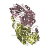
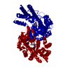
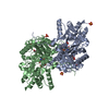
- PDB-2z1y: Crystal structure of LysN, alpha-aminoadipate aminotransferase (c... -
+ Open data

- Basic information
| Entry | Database: PDB / ID: 2z1y | |||||||||
|---|---|---|---|---|---|---|---|---|---|---|
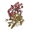
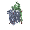
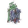
| Title | Crystal structure of LysN, alpha-aminoadipate aminotransferase (complexed with N-(5'-phosphopyridoxyl)-L-leucine), from Thermus thermophilus HB27 | |||||||||
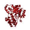
 Components Components | 2-aminoadipate transaminase | |||||||||
 Keywords Keywords | TRANSFERASE / alpha-aminoadipate aminotransferase / thermus thermophilus / substrate specifity | |||||||||
| Function / homology |  Function and homology information Function and homology information2-aminoadipate transaminase / L-2-aminoadipate:2-oxoglutarate transaminase activity / : / pyridoxal phosphate binding Similarity search - Function | |||||||||
| Biological species |   Thermus thermophilus HB27 (bacteria) Thermus thermophilus HB27 (bacteria) | |||||||||
| Method |  X-RAY DIFFRACTION / SYNCHROTRON / MOLECULAR REPLACEMENT / Resolution: 1.75 Å X-RAY DIFFRACTION / SYNCHROTRON / MOLECULAR REPLACEMENT / Resolution: 1.75 Å | |||||||||
 Authors Authors | Tomita, T. / Miyazaki, T. / Miyagawa, T. / Fushinobu, S. / Kuzuyama, T. / Nishiyama, M. | |||||||||
| Funding support | 1items
| |||||||||
 Citation Citation | Journal: To be Published Title: Mechanism of broad substrate specificity of alpha-aminoadipate aminotransferase from Thermus thermophilus Authors: Tomita, T. / Miyazaki, T. / Miyagawa, T. / Fushinobu, S. / Kuzuyama, T. / Nishiyama, M. #1: Journal: Microbiology / Year: 2004 Title: alpha-Aminoadipate aminotransferase from an extremely thermophilic bacterium, Thermus thermophilus Authors: Miyazaki, T. / Miyazaki, J. / Yamane, H. / Nishiyama, M. | |||||||||
| History |
|
- Structure visualization
| Structure viewer | Molecule: MolmilJmol/JSmol |
|---|
- Downloads & links
-Download
| PDBx/mmCIF format | 2z1y.cif.gz | 181.8 KB | Display | PDBx/mmCIF format |
|---|---|---|---|---|
| PDB format | pdb2z1y.ent.gz | 141.5 KB | Display | PDB format |
| PDBx/mmJSON format | 2z1y.json.gz | Tree view | PDBx/mmJSON format | |
| Others |  Other downloads Other downloads |
-Validation report
| Arichive directory | https://data.pdbj.org/pub/pdb/validation_reports/z1/2z1yftp://data.pdbj.org/pub/pdb/validation_reports/z1/2z1y | HTTPS FTP |
|---|
-Related structure data
| Related structure data |  2dtv S: Starting model for refinement |
|---|---|
| Similar structure data |


-Links
 PDBj
PDBj


- Assembly
| Deposited unit | 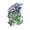
| ||||||||
|---|---|---|---|---|---|---|---|---|---|
| 1 |
| ||||||||
| Unit cell |
|
-Components
| #1: Protein | Mass: 43899.738 Da / Num. of mol.: 2 Source method: isolated from a genetically manipulated source Source: (gene. exp.) Thermus thermophilus HB27 (bacteria) / Gene: lysN, TT_C0043 / Plasmid: pET-LysN7 / Production host: #2: Chemical |   Type: L-peptide linking / Mass: 131.173 Da / Num. of mol.: 2 / Source method: obtained synthetically / Formula: C6H13NO2 Type: L-peptide linking / Mass: 131.173 Da / Num. of mol.: 2 / Source method: obtained synthetically / Formula: C6H13NO2#3: Chemical |   Mass: 247.142 Da / Num. of mol.: 2 / Source method: obtained synthetically / Formula: C8H10NO6P Mass: 247.142 Da / Num. of mol.: 2 / Source method: obtained synthetically / Formula: C8H10NO6P#4: Water | ChemComp-HOH / |  Mass: 18.015 Da / Num. of mol.: 754 / Source method: isolated from a natural source / Formula: H2O Mass: 18.015 Da / Num. of mol.: 754 / Source method: isolated from a natural source / Formula: H2OHas ligand of interest | Y | Has protein modification | N | |
|---|
-Experimental details
-Experiment
| Experiment | Method: X-RAY DIFFRACTION / Number of used crystals: 1 |
|---|
- Sample preparation
| Crystal | Density Matthews: 2.34 Å3/Da / Density % sol: 47.38 % |
|---|---|
| Crystal grow | Temperature: 293 K / Method: vapor diffusion, hanging drop / pH: 7 Details: 18% PEG 4000, 0.1M Tris-HCl (pH 7.0), 0.2M lithium sulfate, VAPOR DIFFUSION, HANGING DROP, temperature 293K |
-Data collection
| Diffraction | Mean temperature: 100 K / Serial crystal experiment: N |
|---|---|
| Diffraction source | Source: SYNCHROTRON / Site: Photon Factory  / Beamline: BL-6A / Wavelength: 0.978 Å / Beamline: BL-6A / Wavelength: 0.978 Å |
| Detector | Type: ADSC QUANTUM 4r / Detector: CCD / Date: Apr 22, 2007 / Details: mirrors |
| Radiation | Monochromator: Si 111 CHANNEL / Protocol: SINGLE WAVELENGTH / Monochromatic (M) / Laue (L): M / Scattering type: x-ray |
| Radiation wavelength | Wavelength: 0.978 Å / Relative weight: 1 |
| Reflection | Resolution: 1.75→50 Å / Num. all: 81957 / Num. obs: 81957 / % possible obs: 100 % / Observed criterion σ(F): 2 / Observed criterion σ(I): 2 / Redundancy: 3.5 % / Biso Wilson estimate: 15.6 Å2 / Rmerge(I) obs: 0.109 / Rsym value: 0.109 / Net I/σ(I): 13.8 |
| Reflection shell | Resolution: 1.75→1.81 Å / Redundancy: 3.5 % / Rmerge(I) obs: 0.323 / Mean I/σ(I) obs: 2.8 / Num. unique obs: 81720 / Rsym value: 0.323 / % possible all: 100 |
- Processing
| Software |
| ||||||||||||||||||||||||||||||||||||||||||||||||||||||||||||||||||||||||||||||||||||||||||||||||||||||||||||||||||||||||||||||||||||||||||||||||||||||||||||||||||||||||||||||||||||||
|---|---|---|---|---|---|---|---|---|---|---|---|---|---|---|---|---|---|---|---|---|---|---|---|---|---|---|---|---|---|---|---|---|---|---|---|---|---|---|---|---|---|---|---|---|---|---|---|---|---|---|---|---|---|---|---|---|---|---|---|---|---|---|---|---|---|---|---|---|---|---|---|---|---|---|---|---|---|---|---|---|---|---|---|---|---|---|---|---|---|---|---|---|---|---|---|---|---|---|---|---|---|---|---|---|---|---|---|---|---|---|---|---|---|---|---|---|---|---|---|---|---|---|---|---|---|---|---|---|---|---|---|---|---|---|---|---|---|---|---|---|---|---|---|---|---|---|---|---|---|---|---|---|---|---|---|---|---|---|---|---|---|---|---|---|---|---|---|---|---|---|---|---|---|---|---|---|---|---|---|---|---|---|---|
| Refinement | Method to determine structure: MOLECULAR REPLACEMENT Starting model: 2DTV 2dtv Resolution: 1.75→36.86 Å / Cor.coef. Fo:Fc: 0.964 / Cor.coef. Fo:Fc free: 0.945 / SU B: 2.226 / SU ML: 0.071 / Cross valid method: THROUGHOUT / ESU R: 0.102 / ESU R Free: 0.104 / Stereochemistry target values: MAXIMUM LIKELIHOOD / Details: HYDROGENS HAVE BEEN ADDED IN THE RIDING POSITIONS
| ||||||||||||||||||||||||||||||||||||||||||||||||||||||||||||||||||||||||||||||||||||||||||||||||||||||||||||||||||||||||||||||||||||||||||||||||||||||||||||||||||||||||||||||||||||||
| Solvent computation | Ion probe radii: 0.8 Å / Shrinkage radii: 0.8 Å / VDW probe radii: 1.2 Å / Solvent model: MASK | ||||||||||||||||||||||||||||||||||||||||||||||||||||||||||||||||||||||||||||||||||||||||||||||||||||||||||||||||||||||||||||||||||||||||||||||||||||||||||||||||||||||||||||||||||||||
| Displacement parameters | Biso mean: 16.14 Å2
| ||||||||||||||||||||||||||||||||||||||||||||||||||||||||||||||||||||||||||||||||||||||||||||||||||||||||||||||||||||||||||||||||||||||||||||||||||||||||||||||||||||||||||||||||||||||
| Refinement step | Cycle: LAST / Resolution: 1.75→36.86 Å
| ||||||||||||||||||||||||||||||||||||||||||||||||||||||||||||||||||||||||||||||||||||||||||||||||||||||||||||||||||||||||||||||||||||||||||||||||||||||||||||||||||||||||||||||||||||||
| Refine LS restraints |
|