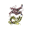
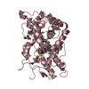
[English] 日本語
Yorodumi- PDB-2yza: Crystal structure of kinase domain of Human 5'-AMP-activated prot... -
+Open data
-Basic information
Entry | Database: PDB / ID: 2yza | ||||||
---|---|---|---|---|---|---|---|
Title | Crystal structure of kinase domain of Human 5'-AMP-activated protein kinase alpha-2 subunit mutant (T172D) | ||||||
Components | 5'-AMP-activated protein kinase catalytic subunit alpha-2 | ||||||
Keywords | TRANSFERASE / Signaling protein / Serine/threonine protein kinase / Phosphorylation / ATP-binding / Nucleotide-binding / Cholesterol biosynthesis / Fatty acid biosynthesis / Lipid synthesis / glucose metabolism / Magnesium / Metal-binding / Serine/threonine-protein kinase / Steroid biosynthesis / Structural Genomics / NPPSFA / National Project on Protein Structural and Functional Analyses / RIKEN Structural Genomics/Proteomics Initiative / RSGI | ||||||
Function / homology | Function and homology information [hydroxymethylglutaryl-CoA reductase (NADPH)] kinase / [hydroxymethylglutaryl-CoA reductase (NADPH)] kinase activity / : / regulation of stress granule assembly / histone H2BS36 kinase activity / AMPK inhibits chREBP transcriptional activation activity / positive regulation of peptidyl-lysine acetylation / lipid droplet disassembly / Lipophagy / nucleotide-activated protein kinase complex ...[hydroxymethylglutaryl-CoA reductase (NADPH)] kinase / [hydroxymethylglutaryl-CoA reductase (NADPH)] kinase activity / : / regulation of stress granule assembly / histone H2BS36 kinase activity / AMPK inhibits chREBP transcriptional activation activity / positive regulation of peptidyl-lysine acetylation / lipid droplet disassembly / Lipophagy / nucleotide-activated protein kinase complex / Energy dependent regulation of mTOR by LKB1-AMPK / Carnitine metabolism / negative regulation of hepatocyte apoptotic process / protein localization to lipid droplet / negative regulation of TOR signaling / response to muscle activity / Activation of PPARGC1A (PGC-1alpha) by phosphorylation / Nuclear events mediated by NFE2L2 / lipid biosynthetic process / AMP-activated protein kinase activity / negative regulation of tubulin deacetylation / Macroautophagy / positive regulation of protein localization / cholesterol biosynthetic process / cellular response to nutrient levels / positive regulation of macroautophagy / regulation of macroautophagy / fatty acid homeostasis / cellular response to glucose starvation / positive regulation of autophagy / energy homeostasis / regulation of microtubule cytoskeleton organization / Activation of AMPK downstream of NMDARs / negative regulation of TORC1 signaling / cellular response to calcium ion / protein serine/threonine/tyrosine kinase activity / positive regulation of glycolytic process / Translocation of SLC2A4 (GLUT4) to the plasma membrane / cellular response to glucose stimulus / TP53 Regulates Metabolic Genes / regulation of circadian rhythm / Wnt signaling pathway / fatty acid biosynthetic process / autophagy / cytoplasmic stress granule / cellular response to prostaglandin E stimulus / rhythmic process / cellular response to xenobiotic stimulus / glucose homeostasis / cellular response to oxidative stress / Regulation of TP53 Activity through Phosphorylation / non-specific serine/threonine protein kinase / protein kinase activity / intracellular signal transduction / nuclear speck / axon / protein phosphorylation / negative regulation of gene expression / protein serine kinase activity / protein serine/threonine kinase activity / neuronal cell body / chromatin binding / dendrite / negative regulation of apoptotic process / Golgi apparatus / signal transduction / nucleoplasm / ATP binding / nucleus / metal ion binding / cytoplasm / cytosol Similarity search - Function | ||||||
Biological species | Homo sapiens (human) | ||||||
Method | X-RAY DIFFRACTION / SYNCHROTRON / MOLECULAR REPLACEMENT / Resolution: 3.02 Å | ||||||
Authors | Saijo, S. / Takagi, T. / Yoshikawa, S. / Kishishita, S. / Shirouzu, M. / Yokoyama, S. / RIKEN Structural Genomics/Proteomics Initiative (RSGI) | ||||||
Citation | Journal: Acta Crystallogr.,Sect.D / Year: 2011 Title: Structural basis for compound C inhibition of the human AMP-activated protein kinase alpha 2 subunit kinase domain Authors: Handa, N. / Takagi, T. / Saijo, S. / Kishishita, S. / Takaya, D. / Toyama, M. / Terada, T. / Shirouzu, M. / Suzuki, A. / Lee, S. / Yamauchi, T. / Okada-Iwabu, M. / Iwabu, M. / Kadowaki, T. / ...Authors: Handa, N. / Takagi, T. / Saijo, S. / Kishishita, S. / Takaya, D. / Toyama, M. / Terada, T. / Shirouzu, M. / Suzuki, A. / Lee, S. / Yamauchi, T. / Okada-Iwabu, M. / Iwabu, M. / Kadowaki, T. / Minokoshi, Y. / Yokoyama, S. | ||||||
History |
|
-Structure visualization
Structure viewer | Molecule: MolmilJmol/JSmol |
---|
-Downloads & links
-Download
PDBx/mmCIF format | 2yza.cif.gz | 62.9 KB | Display | PDBx/mmCIF format |
---|---|---|---|---|
PDB format | pdb2yza.ent.gz | 45.3 KB | Display | PDB format |
PDBx/mmJSON format | 2yza.json.gz | Tree view | PDBx/mmJSON format | |
Others | Other downloads |
-Validation report
Summary document | 2yza_validation.pdf.gz | 423 KB | Display | wwPDB validaton report |
---|---|---|---|---|
Full document | 2yza_full_validation.pdf.gz | 424.3 KB | Display | |
Data in XML | 2yza_validation.xml.gz | 10.6 KB | Display | |
Data in CIF | 2yza_validation.cif.gz | 13.5 KB | Display | |
Arichive directory | https://data.pdbj.org/pub/pdb/validation_reports/yz/2yza ftp://data.pdbj.org/pub/pdb/validation_reports/yz/2yza | HTTPS FTP |
-Related structure data
Related structure data | 3aqvC 2h6dS S: Starting model for refinement C: citing same article (ref.) |
---|---|
Similar structure data |
-Links
-Assembly
Deposited unit |
| ||||||||
---|---|---|---|---|---|---|---|---|---|
1 |
| ||||||||
Unit cell |
|
-Components
#1: Protein | Mass: 31571.730 Da / Num. of mol.: 1 / Fragment: kinase domain / Mutation: T172D Source method: isolated from a genetically manipulated source Source: (gene. exp.) Homo sapiens (human) / Gene: PRKAA2, AMPK, AMPK2 / Plasmid: PX060824-01 / Production host: Cell-free protein synthesis References: UniProt: P54646, non-specific serine/threonine protein kinase |
---|---|
#2: Water | ChemComp-HOH / |
-Experimental details
-Experiment
Experiment | Method: X-RAY DIFFRACTION / Number of used crystals: 1 |
---|
-Sample preparation
Crystal | Density Matthews: 2.11 Å3/Da / Density % sol: 41.82 % |
---|---|
Crystal grow | Temperature: 293 K / Method: vapor diffusion, hanging drop / pH: 8.9 Details: 0.1M Tris-HCl pH8.9, 16% PEG4000, 15% isopropanol, 0.1M Ammonium sulfate, Protein solution: 5mM AMPPNP, 5mM Magnesium chloride, VAPOR DIFFUSION, HANGING DROP, temperature 293K |
-Data collection
Diffraction | Mean temperature: 100 K |
---|---|
Diffraction source | Source: SYNCHROTRON / Site: SPring-8 / Beamline: BL26B2 / Wavelength: 1 Å |
Detector | Type: RIGAKU JUPITER 210 / Detector: CCD / Date: Sep 28, 2006 / Details: rhodium coated mirror |
Radiation | Monochromator: fixed exit Si double crystal monochromator / Protocol: SINGLE WAVELENGTH / Monochromatic (M) / Laue (L): M / Scattering type: x-ray |
Radiation wavelength | Wavelength: 1 Å / Relative weight: 1 |
Reflection | Resolution: 3→25 Å / Num. obs: 4913 / % possible obs: 94.3 % / Redundancy: 3 % / Biso Wilson estimate: 48.3 Å2 / Rsym value: 0.106 / Net I/σ(I): 11.5 |
Reflection shell | Resolution: 3→3.05 Å / Redundancy: 1.7 % / Mean I/σ(I) obs: 3.2 / Num. unique all: 126 / Rsym value: 0.251 / % possible all: 51.6 |
-Processing
Software |
| ||||||||||||||||||||||||||||||||||||||||||||||||||||||||||||||||||||||||||||||||||||||||||||||||||||||||||||||||||||||||||||||||||||||||||||||||||||||||||||||||||||||||||
---|---|---|---|---|---|---|---|---|---|---|---|---|---|---|---|---|---|---|---|---|---|---|---|---|---|---|---|---|---|---|---|---|---|---|---|---|---|---|---|---|---|---|---|---|---|---|---|---|---|---|---|---|---|---|---|---|---|---|---|---|---|---|---|---|---|---|---|---|---|---|---|---|---|---|---|---|---|---|---|---|---|---|---|---|---|---|---|---|---|---|---|---|---|---|---|---|---|---|---|---|---|---|---|---|---|---|---|---|---|---|---|---|---|---|---|---|---|---|---|---|---|---|---|---|---|---|---|---|---|---|---|---|---|---|---|---|---|---|---|---|---|---|---|---|---|---|---|---|---|---|---|---|---|---|---|---|---|---|---|---|---|---|---|---|---|---|---|---|---|---|---|
Refinement | Method to determine structure: MOLECULAR REPLACEMENT Starting model: PDB ENTRY 2H6D Resolution: 3.02→25 Å / Cor.coef. Fo:Fc: 0.887 / Cor.coef. Fo:Fc free: 0.769 / SU B: 25.942 / SU ML: 0.485 / Cross valid method: THROUGHOUT / ESU R Free: 0.673 / Stereochemistry target values: MAXIMUM LIKELIHOOD
| ||||||||||||||||||||||||||||||||||||||||||||||||||||||||||||||||||||||||||||||||||||||||||||||||||||||||||||||||||||||||||||||||||||||||||||||||||||||||||||||||||||||||||
Solvent computation | Ion probe radii: 0.8 Å / Shrinkage radii: 0.8 Å / VDW probe radii: 1.4 Å / Solvent model: MASK | ||||||||||||||||||||||||||||||||||||||||||||||||||||||||||||||||||||||||||||||||||||||||||||||||||||||||||||||||||||||||||||||||||||||||||||||||||||||||||||||||||||||||||
Displacement parameters | Biso mean: 21.753 Å2
| ||||||||||||||||||||||||||||||||||||||||||||||||||||||||||||||||||||||||||||||||||||||||||||||||||||||||||||||||||||||||||||||||||||||||||||||||||||||||||||||||||||||||||
Refinement step | Cycle: LAST / Resolution: 3.02→25 Å
| ||||||||||||||||||||||||||||||||||||||||||||||||||||||||||||||||||||||||||||||||||||||||||||||||||||||||||||||||||||||||||||||||||||||||||||||||||||||||||||||||||||||||||
Refine LS restraints |
| ||||||||||||||||||||||||||||||||||||||||||||||||||||||||||||||||||||||||||||||||||||||||||||||||||||||||||||||||||||||||||||||||||||||||||||||||||||||||||||||||||||||||||
LS refinement shell | Resolution: 3.02→3.094 Å / Total num. of bins used: 20
|