 Movie
Movie Controller


+ Open data

- Basic information
| Entry | Database: PDB / ID: 2yyh | ||||||
|---|---|---|---|---|---|---|---|
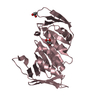
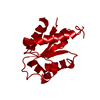
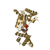
| Title | Crystal structure of Nudix family protein from Aquifex aeolicus | ||||||
 Components Components | 8-OXO-dGTPase domain | ||||||
 Keywords Keywords | HYDROLASE / Nudix family protein / Structural Genomics / NPPSFA / National Project on Protein Structural and Functional Analyses / RIKEN Structural Genomics/Proteomics Initiative / RSGI | ||||||
| Function / homology |  Function and homology information Function and homology informationdihydroneopterin triphosphate pyrophosphohydrolase activity / dATP diphosphatase activity / folic acid biosynthetic process / tetrahydrofolate biosynthetic process Similarity search - Function | ||||||
| Biological species |   Aquifex aeolicus (bacteria) Aquifex aeolicus (bacteria) | ||||||
| Method |  X-RAY DIFFRACTION / SYNCHROTRON / MAD / Resolution: 1.8 Å X-RAY DIFFRACTION / SYNCHROTRON / MAD / Resolution: 1.8 Å | ||||||
 Authors Authors | Nakagawa, N. / Kishishita, S. / Yokoyama, S. / Kuramitsu, S. / RIKEN Structural Genomics/Proteomics Initiative (RSGI) | ||||||
 Citation Citation | Journal: To be Published Title: Crystal structure of Nudix family protein from Aquifex aeolicus Authors: Nakakaga, N. / Kishishita, S. / Yokoyama, S. / Kuramitsu, S. | ||||||
| History |
|
- Structure visualization
| Structure viewer | Molecule: MolmilJmol/JSmol |
|---|
- Downloads & links
-Download
| PDBx/mmCIF format | 2yyh.cif.gz | 121.5 KB | Display | PDBx/mmCIF format |
|---|---|---|---|---|
| PDB format | pdb2yyh.ent.gz | 94.7 KB | Display | PDB format |
| PDBx/mmJSON format | 2yyh.json.gz | Tree view | PDBx/mmJSON format | |
| Others |  Other downloads Other downloads |
-Validation report
| Summary document | 2yyh_validation.pdf.gz | 438.2 KB | Display | wwPDB validaton report |
|---|---|---|---|---|
| Full document | 2yyh_full_validation.pdf.gz | 447 KB | Display | |
| Data in XML | 2yyh_validation.xml.gz | 31.2 KB | Display | |
| Data in CIF | 2yyh_validation.cif.gz | 43 KB | Display | |
| Arichive directory | https://data.pdbj.org/pub/pdb/validation_reports/yy/2yyhftp://data.pdbj.org/pub/pdb/validation_reports/yy/2yyh | HTTPS FTP |
-Related structure data
| Similar structure data | |
|---|---|
| Other databases |


-Links
 PDBj
PDBj

- Assembly
| Deposited unit | 
| ||||||||
|---|---|---|---|---|---|---|---|---|---|
| 1 | 
| ||||||||
| 2 | 
| ||||||||
| Unit cell |
| ||||||||
| Components on special symmetry positions |
|
-Components
| #1: Protein | Mass: 15804.840 Da / Num. of mol.: 4 Source method: isolated from a genetically manipulated source Source: (gene. exp.) Aquifex aeolicus (bacteria) / Strain: VF5 / Gene: mutT / Plasmid: pET-21a / Production host: #2: Water | ChemComp-HOH / |  Mass: 18.015 Da / Num. of mol.: 522 / Source method: isolated from a natural source / Formula: H2O Mass: 18.015 Da / Num. of mol.: 522 / Source method: isolated from a natural source / Formula: H2OHas protein modification | Y | |
|---|
-Experimental details
-Experiment
| Experiment | Method: X-RAY DIFFRACTION / Number of used crystals: 1 |
|---|
- Sample preparation
| Crystal | Density Matthews: 2.07 Å3/Da / Density % sol: 40.54 % / Description: The file contains Friedel pairs. |
|---|---|
| Crystal grow | Temperature: 293 K / Method: vapor diffusion, sitting drop / pH: 6.7 Details: 0.16M LiCl, 16% PEG3350, pH 6.7, VAPOR DIFFUSION, SITTING DROP, temperature 293K |
-Data collection
| Diffraction | Mean temperature: 100 K | ||||||||||||
|---|---|---|---|---|---|---|---|---|---|---|---|---|---|
| Diffraction source | Source: SYNCHROTRON / Site: SPring-8  / Beamline: BL26B2 / Wavelength: 0.97894, 0.9000, 0.97935 / Beamline: BL26B2 / Wavelength: 0.97894, 0.9000, 0.97935 | ||||||||||||
| Detector | Type: RIGAKU JUPITER 210 / Detector: CCD / Date: May 16, 2006 / Details: mirrors | ||||||||||||
| Radiation | Monochromator: Si II / Protocol: MAD / Monochromatic (M) / Laue (L): M / Scattering type: x-ray | ||||||||||||
| Radiation wavelength |
| ||||||||||||
| Reflection | Resolution: 1.8→50 Å / Num. obs: 91555 / % possible obs: 98.5 % / Observed criterion σ(I): -3 / Redundancy: 2.75 % / Biso Wilson estimate: 13.6 Å2 / Rsym value: 0.042 / Net I/σ(I): 18 | ||||||||||||
| Reflection shell | Resolution: 1.8→1.86 Å / Mean I/σ(I) obs: 1.96 / Rsym value: 0.25 / % possible all: 91 |
- Processing
| Software |
| ||||||||||||||||||||||||||||||||||||||||||||||||||||||||||||
|---|---|---|---|---|---|---|---|---|---|---|---|---|---|---|---|---|---|---|---|---|---|---|---|---|---|---|---|---|---|---|---|---|---|---|---|---|---|---|---|---|---|---|---|---|---|---|---|---|---|---|---|---|---|---|---|---|---|---|---|---|---|
| Refinement | Method to determine structure: MAD / Resolution: 1.8→39.5 Å / Rfactor Rfree error: 0.004 / Data cutoff high absF: 389621.31 / Data cutoff low absF: 0 / Isotropic thermal model: RESTRAINED / Cross valid method: THROUGHOUT / σ(F): 0 / Details: the file contains Friedel pairs.
| ||||||||||||||||||||||||||||||||||||||||||||||||||||||||||||
| Solvent computation | Solvent model: FLAT MODEL / Bsol: 50.2637 Å2 / ksol: 0.367316 e/Å3 | ||||||||||||||||||||||||||||||||||||||||||||||||||||||||||||
| Displacement parameters | Biso mean: 24.6 Å2
| ||||||||||||||||||||||||||||||||||||||||||||||||||||||||||||
| Refine analyze |
| ||||||||||||||||||||||||||||||||||||||||||||||||||||||||||||
| Refinement step | Cycle: LAST / Resolution: 1.8→39.5 Å
| ||||||||||||||||||||||||||||||||||||||||||||||||||||||||||||
| Refine LS restraints |
| ||||||||||||||||||||||||||||||||||||||||||||||||||||||||||||
| LS refinement shell | Resolution: 1.8→1.91 Å / Rfactor Rfree error: 0.013 / Total num. of bins used: 6
| ||||||||||||||||||||||||||||||||||||||||||||||||||||||||||||
| Xplor file |
|
