 Movie
Movie Controller


+ Open data

- Basic information
| Entry | Database: PDB / ID: 2yvn | ||||||
|---|---|---|---|---|---|---|---|
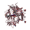
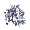
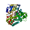
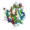
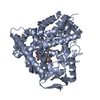
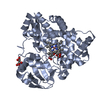
| Title | Crystal structure of NDX2 from thermus thermophilus HB8 | ||||||
 Components Components | MutT/nudix family protein | ||||||
 Keywords Keywords | HYDROLASE / NUDIX PROTEIN / ADP-RIBOSE / FAD / THERMUS THERMOPHILUS / HYDROLAS / Structural Genomics / NPPSFA / National Project on Protein Structural and Functional Analyses / RIKEN Structural Genomics/Proteomics Initiative / RSGI | ||||||
| Function / homology |  Function and homology information Function and homology informationpyrophosphatase activity / nucleoside phosphate metabolic process / ribose phosphate metabolic process / nucleotide binding / metal ion binding / cytosol Similarity search - Function | ||||||
| Biological species |   Thermus thermophilus (bacteria) Thermus thermophilus (bacteria) | ||||||
| Method |  X-RAY DIFFRACTION / SYNCHROTRON / MOLECULAR REPLACEMENT / Resolution: 1.84 Å X-RAY DIFFRACTION / SYNCHROTRON / MOLECULAR REPLACEMENT / Resolution: 1.84 Å | ||||||
 Authors Authors | Wakamatsu, T. / Nakagawa, N. / Kuramitsu, S. / Yokoyama, S. / Masui, R. / RIKEN Structural Genomics/Proteomics Initiative (RSGI) | ||||||
 Citation Citation | Journal: J.Bacteriol. / Year: 2008 Title: Structural basis for different substrate specificities of two ADP-ribose pyrophosphatases from Thermus thermophilus HB8 Authors: Wakamatsu, T. / Nakagawa, N. / Kuramitsu, S. / Masui, R. | ||||||
| History |
|
- Structure visualization
| Structure viewer | Molecule: MolmilJmol/JSmol |
|---|
- Downloads & links
-Download
| PDBx/mmCIF format | 2yvn.cif.gz | 50 KB | Display | PDBx/mmCIF format |
|---|---|---|---|---|
| PDB format | pdb2yvn.ent.gz | 35.6 KB | Display | PDB format |
| PDBx/mmJSON format | 2yvn.json.gz | Tree view | PDBx/mmJSON format | |
| Others |  Other downloads Other downloads |
-Validation report
| Arichive directory | https://data.pdbj.org/pub/pdb/validation_reports/yv/2yvnftp://data.pdbj.org/pub/pdb/validation_reports/yv/2yvn | HTTPS FTP |
|---|
-Related structure data
| Related structure data |  2yvmSC  2yvoC 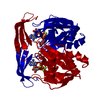 2yvpC S: Starting model for refinement C: citing same article ( |
|---|---|
| Similar structure data | |
| Other databases |


-Links
 PDBj
PDBj

- Assembly
| Deposited unit | 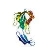
| ||||||||
|---|---|---|---|---|---|---|---|---|---|
| 1 | 
| ||||||||
| Unit cell |
|
-Components
| #1: Protein | Mass: 20336.439 Da / Num. of mol.: 1 Source method: isolated from a genetically manipulated source Source: (gene. exp.) Thermus thermophilus (bacteria) / Strain: HB8 / Gene: ndx2 / Plasmid: pET11a / Species (production host): Escherichia coli / Production host: References: UniProt: Q5SJY9, Hydrolases; Acting on acid anhydrides; In phosphorus-containing anhydrides |
|---|---|
| #2: Water | ChemComp-HOH /  Mass: 18.015 Da / Num. of mol.: 101 / Source method: isolated from a natural source / Formula: H2O Mass: 18.015 Da / Num. of mol.: 101 / Source method: isolated from a natural source / Formula: H2O |
-Experimental details
-Experiment
| Experiment | Method: X-RAY DIFFRACTION / Number of used crystals: 1 |
|---|
- Sample preparation
| Crystal | Density Matthews: 2.5 Å3/Da / Density % sol: 50.73 % |
|---|---|
| Crystal grow | Temperature: 293 K / Method: vapor diffusion, hanging drop / pH: 6.5 Details: 0.05M MES (PH 6.5), 0.08M SODIUM ACETATE, 7%(W/V) PEG 8000, 10%(W/V) GLYCEROL, VAPOR DIFFUSION, HANGING DROP, temperature 293K |
-Data collection
| Diffraction | Mean temperature: 100 K |
|---|---|
| Diffraction source | Source: SYNCHROTRON / Site: SPring-8  / Beamline: BL44B2 / Wavelength: 1 Å / Beamline: BL44B2 / Wavelength: 1 Å |
| Detector | Type: ADSC QUANTUM 210 / Detector: CCD / Date: Jul 29, 2005 |
| Radiation | Monochromator: TRANSPARENT DIAMOND DOUBLE CRYSTAL / Protocol: SINGLE WAVELENGTH / Monochromatic (M) / Laue (L): M / Scattering type: x-ray |
| Radiation wavelength | Wavelength: 1 Å / Relative weight: 1 |
| Reflection | Resolution: 1.79→50 Å / Num. all: 19698 / Num. obs: 19698 / % possible obs: 99.8 % / Observed criterion σ(I): -3 / Redundancy: 5.9 % / Biso Wilson estimate: 9.6 Å2 / Rmerge(I) obs: 0.045 / Net I/σ(I): 37.2 |
| Reflection shell | Resolution: 1.79→1.85 Å / Redundancy: 5.5 % / Rmerge(I) obs: 0.248 / Mean I/σ(I) obs: 5.8 / Num. unique all: 1931 / % possible all: 98.9 |
- Processing
| Software |
| |||||||||||||||||||||||||||
|---|---|---|---|---|---|---|---|---|---|---|---|---|---|---|---|---|---|---|---|---|---|---|---|---|---|---|---|---|
| Refinement | Method to determine structure: MOLECULAR REPLACEMENT Starting model: PDB ENTRY 2YVM Resolution: 1.84→27.36 Å / Isotropic thermal model: RESTRAINED / Cross valid method: THROUGHOUT / σ(F): 0 / Stereochemistry target values: Engh & Huber
| |||||||||||||||||||||||||||
| Displacement parameters | Biso mean: 20.7 Å2
| |||||||||||||||||||||||||||
| Refine analyze |
| |||||||||||||||||||||||||||
| Refinement step | Cycle: LAST / Resolution: 1.84→27.36 Å
| |||||||||||||||||||||||||||
| Refine LS restraints |
| |||||||||||||||||||||||||||
| LS refinement shell | Resolution: 1.84→1.96 Å / Rfactor Rfree error: 0.017
|
