[English] 日本語
Yorodumi
- PDB-2yn3: Structural insight into the giant calcium-binding adhesin SiiE: i... -

+
Open data


ID or keywords:

Loading...

-
Basic information

Entry
Database: PDB / ID: 2yn3
TitleStructural insight into the giant calcium-binding adhesin SiiE: implications for the adhesion of Salmonella enterica to polarized epithelial cells
ComponentsPUTATIVE INNER MEMBRANE PROTEIN
KeywordsMEMBRANE PROTEIN / BIG-DOMAINS ADHESIN
Function / homology
Function and homology information


homophilic cell adhesion via plasma membrane adhesion molecules / calcium ion binding / membrane
Similarity search - Function
N-terminal domain of TfIIb - #90 / Bacterial Ig-like domain / Bacterial Ig-like domain 13 / Bacterial Ig-like domain / Ig-like domain, bacterial type / N-terminal domain of TfIIb / Cadherins domain profile. / Cadherin-like / Other non-globular / Special ...N-terminal domain of TfIIb - #90 / Bacterial Ig-like domain / Bacterial Ig-like domain 13 / Bacterial Ig-like domain / Ig-like domain, bacterial type / N-terminal domain of TfIIb / Cadherins domain profile. / Cadherin-like / Other non-globular / Special / Immunoglobulin E-set / Immunoglobulins / Immunoglobulin-like fold / Immunoglobulin-like / Sandwich / Mainly Beta
Similarity search - Domain/homology
IODIDE ION / PHOSPHATE ION / Inner membrane protein
Similarity search - Component
Biological speciesSALMONELLA ENTERICA SUBSP. ENTERICA SEROVAR TYPHIMURIUM (bacteria)
MethodX-RAY DIFFRACTION / SYNCHROTRON / SAD / Resolution: 2.12 Å
AuthorsGriessl, M.H. / Schmid, B. / Kassler, K. / Braunsmann, C. / Ritter, R. / Barlag, B. / Sturm, K.U. / Danzer, C. / Wagner, C. / Schaeffer, T.E. ...Griessl, M.H. / Schmid, B. / Kassler, K. / Braunsmann, C. / Ritter, R. / Barlag, B. / Sturm, K.U. / Danzer, C. / Wagner, C. / Schaeffer, T.E. / Sticht, H. / Hensel, M. / Muller, Y.A.
Citation
Journal: Structure / Year: 2013
Title: Structural Insight Into the Giant Ca(2+)-Binding Adhesin Siie: Implications for the Adhesion of Salmonella Enterica to Polarized Epithelial Cells.
Authors: Griessl, M.H. / Schmid, B. / Kassler, K. / Braunsmann, C. / Ritter, R. / Barlag, B. / Stierhof, Y. / Sturm, K.U. / Danzer, C. / Wagner, C. / Schaffer, T.E. / Sticht, H. / Hensel, M. / Muller, Y.A.
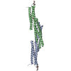
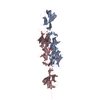
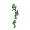
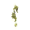
#1: Journal: Acta Crystallogr.,Sect.F / Year: 2011
Title: Crystallization and Preliminary Crystallographic Analysis of an Ig-Domain-Encompassing Fragment of the Giant Adhesion Protein Siie from Salmonella Enterica.
Authors: Sturm, K.U. / Griessl, M.H. / Wagner, C. / Deiwick, J. / Hensel, M. / Muller, Y.A.
History
DepositionOct 11, 2012Deposition site: PDBE / Processing site: PDBE
Revision 1.0Apr 3, 2013Provider: repository / Type: Initial release
Revision 1.1Jun 5, 2013Group: Database references

-
Structure visualization

Structure viewerMolecule:
MolmilJmol/JSmol

Downloads & links

-
Assembly

Deposited unit
A: PUTATIVE INNER MEMBRANE PROTEIN
B: PUTATIVE INNER MEMBRANE PROTEIN
C: PUTATIVE INNER MEMBRANE PROTEIN
D: PUTATIVE INNER MEMBRANE PROTEIN
hetero molecules


Theoretical massNumber of molelcules
Total (without water)122,55720
Polymers121,6874
Non-polymers87016
Water8,323462
1
A: PUTATIVE INNER MEMBRANE PROTEIN
C: PUTATIVE INNER MEMBRANE PROTEIN
hetero molecules


Theoretical massNumber of molelcules
Total (without water)61,47312
Polymers60,8432
Non-polymers62910
Water362
TypeNameSymmetry operationNumber
identity operation1_555x,y,z1
Buried area4720 Å2
ΔGint-107.9 kcal/mol
Surface area26180 Å2
MethodPISA
2
B: PUTATIVE INNER MEMBRANE PROTEIN
hetero molecules


Theoretical massNumber of molelcules
Total (without water)30,5424
Polymers30,4221
Non-polymers1203
Water181
TypeNameSymmetry operationNumber
identity operation1_555x,y,z1
MethodPISA
3
D: PUTATIVE INNER MEMBRANE PROTEIN
hetero molecules


Theoretical massNumber of molelcules
Total (without water)30,5424
Polymers30,4221
Non-polymers1203
Water181
TypeNameSymmetry operationNumber
identity operation1_555x,y,z1
MethodPISA
Unit cell
Length a, b, c (Å)93.480, 153.309, 96.300
Angle α, β, γ (deg.)90.00, 103.88, 90.00
Int Tables number5
Space group name H-MC121

-
Components

#1: Protein
PUTATIVE INNER MEMBRANE PROTEIN / SIIE


Mass: 30421.697 Da / Num. of mol.: 4 / Fragment: BIG DOMAINS 50 TO 52, RESIDUES 5078-5365
Source method: isolated from a genetically manipulated source
Source: (gene. exp.) SALMONELLA ENTERICA SUBSP. ENTERICA SEROVAR TYPHIMURIUM (bacteria)
Strain: LT2 / Plasmid: P3355-1 / Production host: ESCHERICHIA COLI (E. coli) / Strain (production host): BL21(DE3) / References: UniProt: Q8ZKG6
#2: Chemical ChemComp-IOD / IODIDE ION


Mass: 126.904 Da / Num. of mol.: 2 / Source method: obtained synthetically / Formula: I
#3: Chemical
ChemComp-CA / CALCIUM ION


Mass: 40.078 Da / Num. of mol.: 13 / Source method: obtained synthetically / Formula: Ca
#4: Chemical ChemComp-PO4 / PHOSPHATE ION


Mass: 94.971 Da / Num. of mol.: 1 / Source method: obtained synthetically / Formula: PO4
#5: Water ChemComp-HOH / water


Mass: 18.015 Da / Num. of mol.: 462 / Source method: isolated from a natural source / Formula: H2O

-
Experimental details

-
Experiment

ExperimentMethod: X-RAY DIFFRACTION / Number of used crystals: 1

-
Sample preparation

CrystalDensity Matthews: 2.9 Å3/Da / Density % sol: 57 % / Description: NONE
Crystal growDetails: 0.2 M AMMONIUM IODIDE, 20 % PEG 3350

-
Data collection

DiffractionMean temperature: 100 K
Diffraction sourceSource: SYNCHROTRON / Site: BESSY / Beamline: 14.1 / Wavelength: 0.9797
DetectorType: MARRESEARCH / Detector: CCD / Date: Oct 8, 2010 / Details: MIRRORS
RadiationMonochromator: SI-111 CRYSTAL / Protocol: SINGLE WAVELENGTH / Monochromatic (M) / Laue (L): M / Scattering type: x-ray
Radiation wavelengthWavelength: 0.9797 Å / Relative weight: 1
ReflectionResolution: 2.12→35 Å / Num. obs: 72931 / % possible obs: 98 % / Observed criterion σ(I): 2 / Redundancy: 3.7 % / Rmerge(I) obs: 0.076 / Net I/σ(I): 10.7
Reflection shellResolution: 2.12→2.18 Å / Redundancy: 3.1 % / Rmerge(I) obs: 0.396 / Mean I/σ(I) obs: 2.9 / % possible all: 85.6

-
Processing

Software
NameVersionClassification
REFMAC5.7.0029refinement
XDSdata reduction
XSCALEdata scaling
SHELXphasing
RefinementMethod to determine structure: SAD
Starting model: NONE

Resolution: 2.12→34.32 Å / Cor.coef. Fo:Fc: 0.94 / Cor.coef. Fo:Fc free: 0.908 / SU B: 4.757 / SU ML: 0.128 / Cross valid method: THROUGHOUT / ESU R: 0.211 / ESU R Free: 0.191 / Stereochemistry target values: MAXIMUM LIKELIHOOD / Details: HYDROGENS HAVE BEEN ADDED IN THE RIDING POSITIONS.
RfactorNum. reflection% reflectionSelection details
Rfree0.25971 3648 5 %RANDOM
Rwork0.21024 ---
obs0.21271 69306 98.04 %-
Solvent computationIon probe radii: 0.8 Å / Shrinkage radii: 0.8 Å / VDW probe radii: 1.2 Å / Solvent model: MASK
Displacement parametersBiso mean: 41.779 Å2
Baniso -1Baniso -2Baniso -3
1-0.16 Å2-0 Å21.31 Å2
2---1.08 Å20 Å2
3---0.53 Å2
Refinement stepCycle: LAST / Resolution: 2.12→34.32 Å
ProteinNucleic acidLigandSolventTotal
Num. atoms7506 0 20 462 7988
Refine LS restraints
Refine-IDTypeDev idealDev ideal targetNumber
X-RAY DIFFRACTIONr_bond_refined_d0.0120.027640
X-RAY DIFFRACTIONr_bond_other_d
X-RAY DIFFRACTIONr_angle_refined_deg1.5531.95310499
X-RAY DIFFRACTIONr_angle_other_deg
X-RAY DIFFRACTIONr_dihedral_angle_1_deg6.4925999
X-RAY DIFFRACTIONr_dihedral_angle_2_deg38.32527.083312
X-RAY DIFFRACTIONr_dihedral_angle_3_deg14.877151103
X-RAY DIFFRACTIONr_dihedral_angle_4_deg15.8021512
X-RAY DIFFRACTIONr_chiral_restr0.1020.21305
X-RAY DIFFRACTIONr_gen_planes_refined0.0070.0215750
X-RAY DIFFRACTIONr_gen_planes_other
X-RAY DIFFRACTIONr_nbd_refined
X-RAY DIFFRACTIONr_nbd_other
X-RAY DIFFRACTIONr_nbtor_refined
X-RAY DIFFRACTIONr_nbtor_other
X-RAY DIFFRACTIONr_xyhbond_nbd_refined
X-RAY DIFFRACTIONr_xyhbond_nbd_other
X-RAY DIFFRACTIONr_metal_ion_refined
X-RAY DIFFRACTIONr_metal_ion_other
X-RAY DIFFRACTIONr_symmetry_vdw_refined
X-RAY DIFFRACTIONr_symmetry_vdw_other
X-RAY DIFFRACTIONr_symmetry_hbond_refined
X-RAY DIFFRACTIONr_symmetry_hbond_other
X-RAY DIFFRACTIONr_symmetry_metal_ion_refined
X-RAY DIFFRACTIONr_symmetry_metal_ion_other
X-RAY DIFFRACTIONr_mcbond_it
X-RAY DIFFRACTIONr_mcbond_other
X-RAY DIFFRACTIONr_mcangle_it
X-RAY DIFFRACTIONr_mcangle_other
X-RAY DIFFRACTIONr_scbond_it
X-RAY DIFFRACTIONr_scbond_other
X-RAY DIFFRACTIONr_scangle_it
X-RAY DIFFRACTIONr_scangle_other
X-RAY DIFFRACTIONr_long_range_B_refined
X-RAY DIFFRACTIONr_long_range_B_other
X-RAY DIFFRACTIONr_rigid_bond_restr
X-RAY DIFFRACTIONr_sphericity_free
X-RAY DIFFRACTIONr_sphericity_bonded
LS refinement shellResolution: 2.12→2.175 Å / Total num. of bins used: 20
RfactorNum. reflection% reflection
Rfree0.289 233 -
Rwork0.241 4435 -
obs--85.7 %

+
About Yorodumi

-
News

-
Feb 9, 2022. New format data for meta-information of EMDB entries

New format data for meta-information of EMDB entries

  • Version 3 of the EMDB header file is now the official format.
  • The previous official version 1.9 will be removed from the archive.

Related info.:EMDB header

External links:wwPDB to switch to version 3 of the EMDB data model

-
Aug 12, 2020. Covid-19 info

Covid-19 info

URL: https://pdbjlvh1.pdbj.org/emnavi/covid19.php

New page: Covid-19 featured information page in EM Navigator.

Related info.:Covid-19 info / Mar 5, 2020. Novel coronavirus structure data

+
Mar 5, 2020. Novel coronavirus structure data

Novel coronavirus structure data

Related info.:Yorodumi Speices / Aug 12, 2020. Covid-19 info

External links:COVID-19 featured content - PDBj / Molecule of the Month (242):Coronavirus Proteases

+
Jan 31, 2019. EMDB accession codes are about to change! (news from PDBe EMDB page)

EMDB accession codes are about to change! (news from PDBe EMDB page)

  • The allocation of 4 digits for EMDB accession codes will soon come to an end. Whilst these codes will remain in use, new EMDB accession codes will include an additional digit and will expand incrementally as the available range of codes is exhausted. The current 4-digit format prefixed with “EMD-” (i.e. EMD-XXXX) will advance to a 5-digit format (i.e. EMD-XXXXX), and so on. It is currently estimated that the 4-digit codes will be depleted around Spring 2019, at which point the 5-digit format will come into force.
  • The EM Navigator/Yorodumi systems omit the EMD- prefix.

Related info.:Q: What is EMD? / ID/Accession-code notation in Yorodumi/EM Navigator

External links:EMDB Accession Codes are Changing Soon! / Contact to PDBj

+
Jul 12, 2017. Major update of PDB

Major update of PDB

  • wwPDB released updated PDB data conforming to the new PDBx/mmCIF dictionary.
  • This is a major update changing the version number from 4 to 5, and with Remediation, in which all the entries are updated.
  • In this update, many items about electron microscopy experimental information are reorganized (e.g. em_software).
  • Now, EM Navigator and Yorodumi are based on the updated data.

External links:wwPDB Remediation / Enriched Model Files Conforming to OneDep Data Standards Now Available in the PDB FTP Archive

-
Yorodumi

Thousand views of thousand structures

  • Yorodumi is a browser for structure data from EMDB, PDB, SASBDB, etc.
  • This page is also the successor to EM Navigator detail page, and also detail information page/front-end page for Omokage search.
  • The word "yorodu" (or yorozu) is an old Japanese word meaning "ten thousand". "mi" (miru) is to see.

Related info.:EMDB / PDB / SASBDB / Comparison of 3 databanks / Yorodumi Search / Aug 31, 2016. New EM Navigator & Yorodumi / Yorodumi Papers / Jmol/JSmol / Function and homology information / Changes in new EM Navigator and Yorodumi

Read more