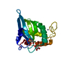
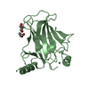
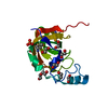
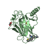
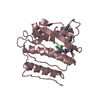
[English] 日本語
Yorodumi
- PDB-2ylf: Crystal structure of the human Spir-1 KIND domain -

+
Open data


ID or keywords:

Loading...

-
Basic information

Entry
Database: PDB / ID: 2ylf
TitleCrystal structure of the human Spir-1 KIND domain
ComponentsPROTEIN SPIRE HOMOLOG 1
KeywordsACTIN-BINDING PROTEIN / ACTIN POLYMERIZATION / SPIR / KINASE
Function / homology
Function and homology information


formin-nucleated actin cable assembly / establishment of meiotic spindle localization / polar body extrusion after meiotic divisions / actin filament network formation / actin nucleation / Golgi vesicle transport / cleavage furrow formation / positive regulation of double-strand break repair / positive regulation of mitochondrial fission / intracellular transport ...formin-nucleated actin cable assembly / establishment of meiotic spindle localization / polar body extrusion after meiotic divisions / actin filament network formation / actin nucleation / Golgi vesicle transport / cleavage furrow formation / positive regulation of double-strand break repair / positive regulation of mitochondrial fission / intracellular transport / vesicle-mediated transport / actin filament polymerization / cytoplasmic vesicle membrane / protein transport / actin binding / cell cortex / actin cytoskeleton organization / mitochondrial outer membrane / cytoskeleton / innate immune response / perinuclear region of cytoplasm / nucleoplasm / plasma membrane / cytosol
Similarity search - Function
Protein Spire1 / Protein Spire / Kinase non-catalytic C-lobe domain / KIND domain / KIND domain profile. / kinase non-catalytic C-lobe domain / Zinc finger, FYVE/PHD-type / Transferase(Phosphotransferase) domain 1 / Transferase(Phosphotransferase); domain 1 / Orthogonal Bundle / Mainly Alpha
Similarity search - Domain/homology
Protein spire homolog 1
Similarity search - Component
Biological speciesHOMO SAPIENS (human)
MethodX-RAY DIFFRACTION / SYNCHROTRON / MOLECULAR REPLACEMENT / Resolution: 2.05 Å
AuthorsZeth, K. / Pechlivanis, M. / Vonrhein, C. / Kerkhoff, E.
CitationJournal: J.Biol.Chem. / Year: 2011
Title: Molecular Basis of Actin Nucleation Factor Cooperativity: Crystal Structure of the Spir-1 Kinase Non-Catalytic C-Lobe Domain (Kind)Formin-2 Formin Spir Interaction Motif (Fsi) Complex.
Authors: Zeth, K. / Pechlivanis, M. / Samol, A. / Pleiser, S. / Vonrhein, C. / Kerkhoff, E.
History
DepositionJun 1, 2011Deposition site: PDBE / Processing site: PDBE
Revision 1.0Jun 8, 2011Provider: repository / Type: Initial release
Revision 1.1Aug 15, 2012Group: Database references / Version format compliance
Revision 1.2Aug 23, 2017Group: Data collection / Category: diffrn_detector / Item: _diffrn_detector.type
Revision 1.3Dec 20, 2023Group: Data collection / Database references ...Data collection / Database references / Other / Refinement description
Category: chem_comp_atom / chem_comp_bond ...chem_comp_atom / chem_comp_bond / database_2 / pdbx_database_status / pdbx_initial_refinement_model
Item: _database_2.pdbx_DOI / _database_2.pdbx_database_accession / _pdbx_database_status.status_code_sf

-
Structure visualization

Structure viewerMolecule:
MolmilJmol/JSmol

Downloads & links

-
Assembly

Deposited unit
A: PROTEIN SPIRE HOMOLOG 1


Theoretical massNumber of molelcules
Total (without water)25,8391
Polymers25,8391
Non-polymers00
Water1,910106
1


  • Idetical with deposited unit
  • defined by author&software
TypeNameSymmetry operationNumber
identity operation1_555x,y,z1
MethodPISA
Unit cell
Length a, b, c (Å)65.914, 65.914, 78.803
Angle α, β, γ (deg.)90.00, 90.00, 120.00
Int Tables number154
Space group name H-MP3221

-
Components

#1: Protein PROTEIN SPIRE HOMOLOG 1 / HUMAN SPIR-1 KIND / SPIR-1


Mass: 25838.787 Da / Num. of mol.: 1 / Fragment: HUMAN SPIR-1 DOMAIN (KIND), RESIDUES 36-236
Source method: isolated from a genetically manipulated source
Source: (gene. exp.) HOMO SAPIENS (human) / Production host: ESCHERICHIA COLI (E. coli) / Strain (production host): BL21(DE3) / Variant (production host): ROSETTA / References: UniProt: Q08AE8
#2: Water ChemComp-HOH / water


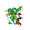
Mass: 18.015 Da / Num. of mol.: 106 / Source method: isolated from a natural source / Formula: H2O

-
Experimental details

-
Experiment

ExperimentMethod: X-RAY DIFFRACTION / Number of used crystals: 1

-
Sample preparation

CrystalDensity Matthews: 1.91 Å3/Da / Density % sol: 35.69 % / Description: NONE
Crystal growpH: 7.5 / Details: pH 7.5

-
Data collection

DiffractionMean temperature: 90 K
Diffraction sourceSource: SYNCHROTRON / Site: SLS / Beamline: X10SA / Wavelength: 1
DetectorType: DECTRIS PILATUS 6M / Detector: PIXEL
RadiationProtocol: SINGLE WAVELENGTH / Monochromatic (M) / Laue (L): M / Scattering type: x-ray
Radiation wavelengthWavelength: 1 Å / Relative weight: 1
ReflectionResolution: 2.05→33 Å / Num. obs: 11995 / % possible obs: 97.5 % / Observed criterion σ(I): 2.8 / Redundancy: 4 % / Rmerge(I) obs: 0.08 / Net I/σ(I): 14.1
Reflection shellResolution: 2.05→2.17 Å / Redundancy: 3.8 % / Rmerge(I) obs: 0.42 / Mean I/σ(I) obs: 2.8 / % possible all: 94.2

-
Processing

Software
NameVersionClassification
REFMAC5.5.0109refinement
XDSdata reduction
XDSdata scaling
MOLREPphasing
RefinementMethod to determine structure: MOLECULAR REPLACEMENT
Starting model: PDB ENTRY 2YLE
Resolution: 2.05→32.96 Å / Cor.coef. Fo:Fc: 0.943 / Cor.coef. Fo:Fc free: 0.909 / SU B: 11.057 / SU ML: 0.134 / Cross valid method: THROUGHOUT / ESU R: 0.197 / ESU R Free: 0.189 / Stereochemistry target values: MAXIMUM LIKELIHOOD / Details: HYDROGENS HAVE BEEN ADDED IN THE RIDING POSITIONS.
RfactorNum. reflection% reflectionSelection details
Rfree0.26339 632 5 %RANDOM
Rwork0.19962 ---
obs0.20283 11995 100 %-
Solvent computationIon probe radii: 0.8 Å / Shrinkage radii: 0.8 Å / VDW probe radii: 1.4 Å / Solvent model: MASK
Displacement parametersBiso mean: 31.341 Å2
Baniso -1Baniso -2Baniso -3
1--0.51 Å2-0.25 Å20 Å2
2---0.51 Å20 Å2
3---0.76 Å2
Refinement stepCycle: LAST / Resolution: 2.05→32.96 Å
ProteinNucleic acidLigandSolventTotal
Num. atoms1300 0 0 106 1406
Refine LS restraints
Refine-IDTypeDev idealDev ideal targetNumber
X-RAY DIFFRACTIONr_bond_refined_d0.0230.0221333
X-RAY DIFFRACTIONr_bond_other_d
X-RAY DIFFRACTIONr_angle_refined_deg1.8071.961804
X-RAY DIFFRACTIONr_angle_other_deg
X-RAY DIFFRACTIONr_dihedral_angle_1_deg6.1065163
X-RAY DIFFRACTIONr_dihedral_angle_2_deg36.57822.83667
X-RAY DIFFRACTIONr_dihedral_angle_3_deg15.13315238
X-RAY DIFFRACTIONr_dihedral_angle_4_deg22.2861516
X-RAY DIFFRACTIONr_chiral_restr0.1310.2201
X-RAY DIFFRACTIONr_gen_planes_refined0.0110.0211012
X-RAY DIFFRACTIONr_gen_planes_other
X-RAY DIFFRACTIONr_nbd_refined
X-RAY DIFFRACTIONr_nbd_other
X-RAY DIFFRACTIONr_nbtor_refined
X-RAY DIFFRACTIONr_nbtor_other
X-RAY DIFFRACTIONr_xyhbond_nbd_refined
X-RAY DIFFRACTIONr_xyhbond_nbd_other
X-RAY DIFFRACTIONr_metal_ion_refined
X-RAY DIFFRACTIONr_metal_ion_other
X-RAY DIFFRACTIONr_symmetry_vdw_refined
X-RAY DIFFRACTIONr_symmetry_vdw_other
X-RAY DIFFRACTIONr_symmetry_hbond_refined
X-RAY DIFFRACTIONr_symmetry_hbond_other
X-RAY DIFFRACTIONr_symmetry_metal_ion_refined
X-RAY DIFFRACTIONr_symmetry_metal_ion_other
X-RAY DIFFRACTIONr_mcbond_it1.2971.5815
X-RAY DIFFRACTIONr_mcbond_other
X-RAY DIFFRACTIONr_mcangle_it2.29221311
X-RAY DIFFRACTIONr_mcangle_other
X-RAY DIFFRACTIONr_scbond_it3.2533518
X-RAY DIFFRACTIONr_scbond_other
X-RAY DIFFRACTIONr_scangle_it5.2384.5491
X-RAY DIFFRACTIONr_scangle_other
X-RAY DIFFRACTIONr_long_range_B_refined
X-RAY DIFFRACTIONr_long_range_B_other
X-RAY DIFFRACTIONr_rigid_bond_restr
X-RAY DIFFRACTIONr_sphericity_free
X-RAY DIFFRACTIONr_sphericity_bonded
LS refinement shellResolution: 2.05→2.103 Å / Total num. of bins used: 20
RfactorNum. reflection% reflection
Rfree0.421 45 -
Rwork0.282 852 -
obs--100 %
Refinement TLS params.Method: refined / Origin x: -28.9336 Å / Origin y: -3.012 Å / Origin z: -8.5915 Å
111213212223313233
T0.0825 Å20.0279 Å2-0.0001 Å2-0.0168 Å20.0014 Å2--0.0608 Å2
L0.5966 °2-0.5764 °20.1408 °2-0.7098 °2-0.1597 °2--0.5898 °2
S-0.0048 Å °-0.0396 Å °0.0467 Å °-0.0066 Å °0.0077 Å °-0.053 Å °-0.0358 Å °-0.017 Å °-0.0028 Å °

+
About Yorodumi

-
News

-
Feb 9, 2022. New format data for meta-information of EMDB entries

New format data for meta-information of EMDB entries

  • Version 3 of the EMDB header file is now the official format.
  • The previous official version 1.9 will be removed from the archive.

Related info.:EMDB header

External links:wwPDB to switch to version 3 of the EMDB data model

-
Aug 12, 2020. Covid-19 info

Covid-19 info

URL: https://pdbjlvh1.pdbj.org/emnavi/covid19.php

New page: Covid-19 featured information page in EM Navigator.

Related info.:Covid-19 info / Mar 5, 2020. Novel coronavirus structure data

+
Mar 5, 2020. Novel coronavirus structure data

Novel coronavirus structure data

Related info.:Yorodumi Speices / Aug 12, 2020. Covid-19 info

External links:COVID-19 featured content - PDBj / Molecule of the Month (242):Coronavirus Proteases

+
Jan 31, 2019. EMDB accession codes are about to change! (news from PDBe EMDB page)

EMDB accession codes are about to change! (news from PDBe EMDB page)

  • The allocation of 4 digits for EMDB accession codes will soon come to an end. Whilst these codes will remain in use, new EMDB accession codes will include an additional digit and will expand incrementally as the available range of codes is exhausted. The current 4-digit format prefixed with “EMD-” (i.e. EMD-XXXX) will advance to a 5-digit format (i.e. EMD-XXXXX), and so on. It is currently estimated that the 4-digit codes will be depleted around Spring 2019, at which point the 5-digit format will come into force.
  • The EM Navigator/Yorodumi systems omit the EMD- prefix.

Related info.:Q: What is EMD? / ID/Accession-code notation in Yorodumi/EM Navigator

External links:EMDB Accession Codes are Changing Soon! / Contact to PDBj

+
Jul 12, 2017. Major update of PDB

Major update of PDB

  • wwPDB released updated PDB data conforming to the new PDBx/mmCIF dictionary.
  • This is a major update changing the version number from 4 to 5, and with Remediation, in which all the entries are updated.
  • In this update, many items about electron microscopy experimental information are reorganized (e.g. em_software).
  • Now, EM Navigator and Yorodumi are based on the updated data.

External links:wwPDB Remediation / Enriched Model Files Conforming to OneDep Data Standards Now Available in the PDB FTP Archive

-
Yorodumi

Thousand views of thousand structures

  • Yorodumi is a browser for structure data from EMDB, PDB, SASBDB, etc.
  • This page is also the successor to EM Navigator detail page, and also detail information page/front-end page for Omokage search.
  • The word "yorodu" (or yorozu) is an old Japanese word meaning "ten thousand". "mi" (miru) is to see.

Related info.:EMDB / PDB / SASBDB / Comparison of 3 databanks / Yorodumi Search / Aug 31, 2016. New EM Navigator & Yorodumi / Yorodumi Papers / Jmol/JSmol / Function and homology information / Changes in new EM Navigator and Yorodumi

Read more