 Movie
Movie Controller

[English] 日本語
 Yorodumi
- PDB-2yjq: Structure of a Paenibacillus Polymyxa Xyloglucanase from Glycosid... -
+ Open data

- Basic information
| Entry | Database: PDB / ID: 2yjq | |||||||||
|---|---|---|---|---|---|---|---|---|---|---|
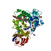
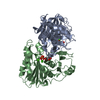
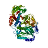
| Title | Structure of a Paenibacillus Polymyxa Xyloglucanase from Glycoside Hydrolase Family 44 | |||||||||
 Components Components | CEL44C | |||||||||
 Keywords Keywords | HYDROLASE / GH44 / ENDO-GLUCANASE / CARBOHYDRATE-BINDING PROTEIN | |||||||||
| Function / homology |  Function and homology information Function and homology informationsubstituted mannan metabolic process / mannan endo-1,4-beta-mannosidase activity / cellulose binding / metal ion binding Similarity search - Function | |||||||||
| Biological species |  PAENIBACILLUS POLYMYXA (bacteria) PAENIBACILLUS POLYMYXA (bacteria) | |||||||||
| Method |  X-RAY DIFFRACTION / SYNCHROTRON / MOLECULAR REPLACEMENT / Resolution: 2.25 Å X-RAY DIFFRACTION / SYNCHROTRON / MOLECULAR REPLACEMENT / Resolution: 2.25 Å | |||||||||
 Authors Authors | Ariza, A. / Eklof, J.M. / Spadiut, O. / Offen, W.A. / Roberts, S.M. / Besenmatter, W. / Friis, E.P. / Skjot, M. / Wilson, K.S. / Brumer, H. / Davies, G. | |||||||||
 Citation Citation | Journal: J.Biol.Chem. / Year: 2011 Title: Structure and Activity of Paenibacillus Polymyxa Xyloglucanase from Glycoside Hydrolase Family 44. Authors: Ariza, A. / Eklof, J.M. / Spadiut, O. / Offen, W.A. / Roberts, S.M. / Besenmatter, W. / Friis, E.P. / Skjot, M. / Wilson, K.S. / Brumer, H. / Davies, G. | |||||||||
| History |
| |||||||||
| Remark 700 | SHEET DETERMINATION METHOD: DSSP THE SHEETS PRESENTED AS "AD" IN EACH CHAIN ON SHEET RECORDS BELOW ... SHEET DETERMINATION METHOD: DSSP THE SHEETS PRESENTED AS "AD" IN EACH CHAIN ON SHEET RECORDS BELOW IS ACTUALLY AN 8-STRANDED BARREL THIS IS REPRESENTED BY A 9-STRANDED SHEET IN WHICH THE FIRST AND LAST STRANDS ARE IDENTICAL. THE SHEETS PRESENTED AS "BD" IN EACH CHAIN ON SHEET RECORDS BELOW IS ACTUALLY AN 8-STRANDED BARREL THIS IS REPRESENTED BY A 9-STRANDED SHEET IN WHICH THE FIRST AND LAST STRANDS ARE IDENTICAL. |
- Structure visualization
| Structure viewer | Molecule: MolmilJmol/JSmol |
|---|
- Downloads & links
-Download
| PDBx/mmCIF format | 2yjq.cif.gz | 223 KB | Display | PDBx/mmCIF format |
|---|---|---|---|---|
| PDB format | pdb2yjq.ent.gz | 177.9 KB | Display | PDB format |
| PDBx/mmJSON format | 2yjq.json.gz | Tree view | PDBx/mmJSON format | |
| Others |  Other downloads Other downloads |
-Validation report
| Arichive directory | https://data.pdbj.org/pub/pdb/validation_reports/yj/2yjqftp://data.pdbj.org/pub/pdb/validation_reports/yj/2yjq | HTTPS FTP |
|---|
-Related structure data
| Related structure data |  2yihC  2ykkSC 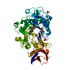 3zq9C C: citing same article ( S: Starting model for refinement |
|---|---|
| Similar structure data |


-Links
 PDBj
PDBj


- Assembly
| Deposited unit | 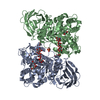
| ||||||||
|---|---|---|---|---|---|---|---|---|---|
| 1 | 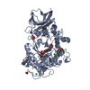
| ||||||||
| 2 | 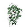
| ||||||||
| Unit cell |
|
-Components
-Protein / Sugars , 2 types, 4 molecules AB
| #1: Protein | Mass: 57915.293 Da / Num. of mol.: 2 / Fragment: RESIDUES 36-559 / Mutation: YES Source method: isolated from a genetically manipulated source Source: (gene. exp.) PAENIBACILLUS POLYMYXA (bacteria) / Strain: GS01 / Production host: References: UniProt: Q1A2D0, cellulase, xyloglucan-specific endo-beta-1,4-glucanase #2: Polysaccharide |   Source method: isolated from a genetically manipulated source Details: oligosaccharide / References: beta-cellobiose |
|---|
-Non-polymers , 7 types, 458 molecules 


| #3: Chemical |  Mass: 149.145 Da / Num. of mol.: 2 / Source method: obtained synthetically / Formula: C5H11NO4 Mass: 149.145 Da / Num. of mol.: 2 / Source method: obtained synthetically / Formula: C5H11NO4#4: Chemical |  Mass: 40.078 Da / Num. of mol.: 2 / Source method: obtained synthetically / Formula: Ca Mass: 40.078 Da / Num. of mol.: 2 / Source method: obtained synthetically / Formula: Ca#5: Chemical | ChemComp-EDO /  Mass: 62.068 Da / Num. of mol.: 10 / Source method: obtained synthetically / Formula: C2H6O2 Mass: 62.068 Da / Num. of mol.: 10 / Source method: obtained synthetically / Formula: C2H6O2#6: Chemical |  Mass: 35.453 Da / Num. of mol.: 2 / Source method: obtained synthetically / Formula: Cl Mass: 35.453 Da / Num. of mol.: 2 / Source method: obtained synthetically / Formula: Cl#7: Chemical |  Mass: 96.063 Da / Num. of mol.: 2 / Source method: obtained synthetically / Formula: SO4 Mass: 96.063 Da / Num. of mol.: 2 / Source method: obtained synthetically / Formula: SO4#8: Chemical | ChemComp-PGE / |  Mass: 150.173 Da / Num. of mol.: 1 / Source method: obtained synthetically / Formula: C6H14O4 Mass: 150.173 Da / Num. of mol.: 1 / Source method: obtained synthetically / Formula: C6H14O4#9: Water | ChemComp-HOH / | Mass: 18.015 Da / Num. of mol.: 439 / Source method: isolated from a natural source / Formula: H2O |
|---|
-Details
| Compound details | ENGINEERED RESIDUE IN CHAIN A, GLN 103 TO HIS ENGINEERED RESIDUE IN CHAIN A, THR 127 TO VAL ...ENGINEERED |
|---|---|
| Has protein modification | Y |
| Sequence details | SEQUENCE IN DATABASE IS SEQUENCE 2 FROM PATENT US 6815192, WHICH IS FOR A XYLOGLUCANASE-BETA- ...SEQUENCE IN DATABASE IS SEQUENCE 2 FROM PATENT US 6815192, WHICH IS FOR A XYLOGLUCAN |
-Experimental details
-Experiment
| Experiment | Method: X-RAY DIFFRACTION / Number of used crystals: 1 |
|---|
- Sample preparation
| Crystal | Density Matthews: 2.8 Å3/Da / Density % sol: 57 % / Description: NONE |
|---|---|
| Crystal grow | Details: 25% (W/V) PEG 3350, 0.2M LITHIUM SULPHATE, 0.1M BIS TRIS PH 6.5 |
-Data collection
| Diffraction | Mean temperature: 120 K |
|---|---|
| Diffraction source | Source: SYNCHROTRON / Site: Diamond  / Beamline: I02 / Wavelength: 0.9795 / Beamline: I02 / Wavelength: 0.9795 |
| Detector | Type: ADSC CCD / Detector: CCD / Date: Oct 7, 2010 |
| Radiation | Protocol: SINGLE WAVELENGTH / Monochromatic (M) / Laue (L): M / Scattering type: x-ray |
| Radiation wavelength | Wavelength: 0.9795 Å / Relative weight: 1 |
| Reflection | Resolution: 2.25→80.01 Å / Num. obs: 62986 / % possible obs: 98.5 % / Observed criterion σ(I): 2 / Redundancy: 8.1 % / Biso Wilson estimate: 35.2 Å2 / Rmerge(I) obs: 0.1 / Net I/σ(I): 14.9 |
| Reflection shell | Resolution: 2.25→2.37 Å / Redundancy: 5.4 % / Rmerge(I) obs: 0.56 / Mean I/σ(I) obs: 2.7 / % possible all: 92.5 |
- Processing
| Software |
| ||||||||||||||||||||||||||||||||||||||||||||||||||||||||||||||||||||||||||||||||||||||||||||||||||||||||||||||||||||||||||||||||||||||||||||||||||||||||||||||||||||||||||||||||||||||
|---|---|---|---|---|---|---|---|---|---|---|---|---|---|---|---|---|---|---|---|---|---|---|---|---|---|---|---|---|---|---|---|---|---|---|---|---|---|---|---|---|---|---|---|---|---|---|---|---|---|---|---|---|---|---|---|---|---|---|---|---|---|---|---|---|---|---|---|---|---|---|---|---|---|---|---|---|---|---|---|---|---|---|---|---|---|---|---|---|---|---|---|---|---|---|---|---|---|---|---|---|---|---|---|---|---|---|---|---|---|---|---|---|---|---|---|---|---|---|---|---|---|---|---|---|---|---|---|---|---|---|---|---|---|---|---|---|---|---|---|---|---|---|---|---|---|---|---|---|---|---|---|---|---|---|---|---|---|---|---|---|---|---|---|---|---|---|---|---|---|---|---|---|---|---|---|---|---|---|---|---|---|---|---|
| Refinement | Method to determine structure: MOLECULAR REPLACEMENT Starting model: APO STRUCTURE, PDB ENTRY 2YKK Resolution: 2.25→106.68 Å / Cor.coef. Fo:Fc: 0.941 / Cor.coef. Fo:Fc free: 0.902 / SU B: 6.396 / SU ML: 0.158 / Cross valid method: THROUGHOUT / ESU R: 0.263 / ESU R Free: 0.226 / Stereochemistry target values: MAXIMUM LIKELIHOOD Details: HYDROGENS HAVE BEEN ADDED IN THE RIDING POSITIONS. RESIDUES 1-6, 442 AND 518-524 IN CHAIN A ARE DISORDERED. RESIDUES 1-7, 442-445 AND 518-524 IN CHAIN B ARE DISORDERED.
| ||||||||||||||||||||||||||||||||||||||||||||||||||||||||||||||||||||||||||||||||||||||||||||||||||||||||||||||||||||||||||||||||||||||||||||||||||||||||||||||||||||||||||||||||||||||
| Solvent computation | Ion probe radii: 0.8 Å / Shrinkage radii: 0.8 Å / VDW probe radii: 1.2 Å / Solvent model: MASK | ||||||||||||||||||||||||||||||||||||||||||||||||||||||||||||||||||||||||||||||||||||||||||||||||||||||||||||||||||||||||||||||||||||||||||||||||||||||||||||||||||||||||||||||||||||||
| Displacement parameters | Biso mean: 29.756 Å2
| ||||||||||||||||||||||||||||||||||||||||||||||||||||||||||||||||||||||||||||||||||||||||||||||||||||||||||||||||||||||||||||||||||||||||||||||||||||||||||||||||||||||||||||||||||||||
| Refinement step | Cycle: LAST / Resolution: 2.25→106.68 Å
| ||||||||||||||||||||||||||||||||||||||||||||||||||||||||||||||||||||||||||||||||||||||||||||||||||||||||||||||||||||||||||||||||||||||||||||||||||||||||||||||||||||||||||||||||||||||
| Refine LS restraints |
|