 Movie
Movie Controller


+ Open data

- Basic information
| Entry | Database: PDB / ID: 4pvq | |||||||||
|---|---|---|---|---|---|---|---|---|---|---|
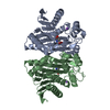
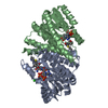
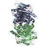
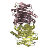
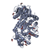
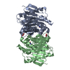
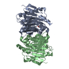
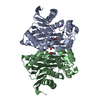
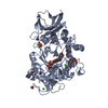
| Title | Crystal structure of sulfate-bound human l-asparaginase protein | |||||||||
 Components Components | Isoaspartyl peptidase/L-asparaginase | |||||||||
 Keywords Keywords | HYDROLASE / NTN ENZYME / HOMODIMER / L-ASPARAGINE | |||||||||
| Function / homology |  Function and homology information Function and homology information: / beta-aspartyl-peptidase / Phenylalanine metabolism / asparaginase / asparaginase activity / beta-aspartyl-peptidase activity / photoreceptor inner segment / proteolysis / nucleus / cytosol / cytoplasm Similarity search - Function | |||||||||
| Biological species |  Homo sapiens (human) Homo sapiens (human) | |||||||||
| Method |  X-RAY DIFFRACTION / MOLECULAR REPLACEMENT / Resolution: 2.13 Å X-RAY DIFFRACTION / MOLECULAR REPLACEMENT / Resolution: 2.13 Å | |||||||||
 Authors Authors | Nomme, J. / Lavie, A. | |||||||||
 Citation Citation | Journal: Biochemistry / Year: 2012 Title: Structures of apo and product-bound human L-asparaginase: insights into the mechanism of autoproteolysis and substrate hydrolysis. Authors: Nomme, J. / Su, Y. / Konrad, M. / Lavie, A. | |||||||||
| History |
|
- Structure visualization
| Structure viewer | Molecule: MolmilJmol/JSmol |
|---|
- Downloads & links
-Download
| PDBx/mmCIF format | 4pvq.cif.gz | 125.9 KB | Display | PDBx/mmCIF format |
|---|---|---|---|---|
| PDB format | pdb4pvq.ent.gz | 96.8 KB | Display | PDB format |
| PDBx/mmJSON format | 4pvq.json.gz | Tree view | PDBx/mmJSON format | |
| Others |  Other downloads Other downloads |
-Validation report
| Arichive directory | https://data.pdbj.org/pub/pdb/validation_reports/pv/4pvqftp://data.pdbj.org/pub/pdb/validation_reports/pv/4pvq | HTTPS FTP |
|---|
-Related structure data
| Related structure data |  4pvpC 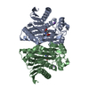 4pvrC  4pvsC  4gdt C: citing same article ( S: Starting model for refinement |
|---|---|
| Similar structure data |

-Links
 PDBj
PDBj


- Assembly
| Deposited unit | 
| ||||||||
|---|---|---|---|---|---|---|---|---|---|
| 1 |
| ||||||||
| Unit cell |
|
-Components
| #1: Protein | Mass: 32289.689 Da / Num. of mol.: 2 Source method: isolated from a genetically manipulated source Source: (gene. exp.) Homo sapiens (human) / Gene: ALP, ASRGL1, CRASH / Plasmid: PET14B / Production host:  References: UniProt: Q7L266, beta-aspartyl-peptidase, asparaginase #2: Chemical | ChemComp-SO4 /   Mass: 96.063 Da / Num. of mol.: 13 / Source method: obtained synthetically / Formula: SO4 Mass: 96.063 Da / Num. of mol.: 13 / Source method: obtained synthetically / Formula: SO4#3: Chemical | ChemComp-IOD /   Mass: 126.904 Da / Num. of mol.: 4 / Source method: obtained synthetically / Formula: I Mass: 126.904 Da / Num. of mol.: 4 / Source method: obtained synthetically / Formula: I#4: Chemical |   Mass: 22.990 Da / Num. of mol.: 2 / Source method: obtained synthetically / Formula: Na Mass: 22.990 Da / Num. of mol.: 2 / Source method: obtained synthetically / Formula: Na#5: Water | ChemComp-HOH / |  Mass: 18.015 Da / Num. of mol.: 212 / Source method: isolated from a natural source / Formula: H2O Mass: 18.015 Da / Num. of mol.: 212 / Source method: isolated from a natural source / Formula: H2O |
|---|
-Experimental details
-Experiment
| Experiment | Method: X-RAY DIFFRACTION / Number of used crystals: 1 |
|---|
- Sample preparation
| Crystal | Density Matthews: 2.25 Å3/Da / Density % sol: 45.26 % |
|---|---|
| Crystal grow | Temperature: 293 K / Method: vapor diffusion, hanging drop / pH: 5.27 Details: 0.2 M AMMONIUM IODIDE, 2.2 M AMMONIUM SULFATE, PH 5.27, VAPOR DIFFUSION, HANGING DROP, TEMPERATURE 293K |
-Data collection
| Diffraction | Mean temperature: 93 K | |||||||||||||||
|---|---|---|---|---|---|---|---|---|---|---|---|---|---|---|---|---|
| Diffraction source | Source: ROTATING ANODE / Type: RIGAKU RU200 / Wavelength: 1.5418 / Wavelength: 1.5418 Å | |||||||||||||||
| Detector | Type: RIGAKU RAXIS IV++ / Detector: IMAGE PLATE / Date: Jan 9, 2012 / Details: MIRRORS | |||||||||||||||
| Radiation | Monochromator: MIRRORS / Protocol: SINGLE WAVELENGTH / Monochromatic (M) / Laue (L): M / Scattering type: x-ray | |||||||||||||||
| Radiation wavelength | Wavelength: 1.5418 Å / Relative weight: 1 | |||||||||||||||
| Reflection twin |
| |||||||||||||||
| Reflection | Resolution: 2.13→30 Å / Num. all: 26285 / Num. obs: 26285 / % possible obs: 79.4 % / Observed criterion σ(F): 0 / Observed criterion σ(I): 0 / Rsym value: 0.067 / Net I/σ(I): 12.8 | |||||||||||||||
| Reflection shell | Resolution: 2.13→2.25 Å / Redundancy: 2.06 % / Mean I/σ(I) obs: 2.89 / Rsym value: 0.325 / % possible all: 53.5 |
- Processing
| Software |
| |||||||||||||||||||||||||||||||||||||||||||||||||||||||||||||||||
|---|---|---|---|---|---|---|---|---|---|---|---|---|---|---|---|---|---|---|---|---|---|---|---|---|---|---|---|---|---|---|---|---|---|---|---|---|---|---|---|---|---|---|---|---|---|---|---|---|---|---|---|---|---|---|---|---|---|---|---|---|---|---|---|---|---|---|
| Refinement | Method to determine structure: MOLECULAR REPLACEMENT Starting model: pdb entry 4GDT 4gdt Resolution: 2.13→30 Å / Cor.coef. Fo:Fc: 0.955 / Cor.coef. Fo:Fc free: 0.935 / SU B: 2.913 / SU ML: 0.08 / Cross valid method: THROUGHOUT / σ(F): 0 / ESU R: 0.071 / ESU R Free: 0.048 / Stereochemistry target values: MAXIMUM LIKELIHOOD / Details: HYDROGENS HAVE BEEN ADDED IN THE RIDING POSITIONS
| |||||||||||||||||||||||||||||||||||||||||||||||||||||||||||||||||
| Solvent computation | Ion probe radii: 0.8 Å / Shrinkage radii: 0.8 Å / VDW probe radii: 1.4 Å / Solvent model: MASK | |||||||||||||||||||||||||||||||||||||||||||||||||||||||||||||||||
| Displacement parameters | Biso mean: 27.2 Å2
| |||||||||||||||||||||||||||||||||||||||||||||||||||||||||||||||||
| Refinement step | Cycle: LAST / Resolution: 2.13→30 Å
| |||||||||||||||||||||||||||||||||||||||||||||||||||||||||||||||||
| Refine LS restraints |
| |||||||||||||||||||||||||||||||||||||||||||||||||||||||||||||||||
| LS refinement shell | Resolution: 2.13→2.18 Å / Total num. of bins used: 20
|
