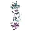
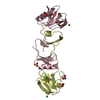
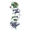
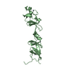
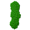
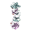
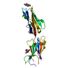
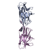
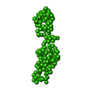
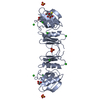
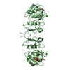
Entry Database : PDB / ID : 2yioTitle Crystal Structure of Parasite Sarcocystis muris Microneme Protein SML- 2 in complex with 1-Thio-beta-D-Galactose (SPACEGROUP C2221) MICRONEME ANTIGEN L2 Keywords / / / / / / Function / homology Function Domain/homology Component
/ / / / / / / / / / / / / / / / / / / / / / Biological species SARCOCYSTIS MURIS (eukaryote)Method / / Resolution : 2.43 Å Authors Mueller, J.J. / Heinemann, U. Journal : Acta Crystallogr.,Sect.D / Year : 2011Title : Pan-Modular Structure of Microneme Protein Sml-2 from Parasite Sarcocystis Muris at 1.95 A Resolution and its Complex with 1-Thio-Beta-D-Galactose.Authors : Mueller, J.J. / Weiss, M.S. / Heinemann, U. History Deposition May 16, 2011 Deposition site / Processing site Revision 1.0 Nov 2, 2011 Provider / Type Revision 1.1 Nov 30, 2011 Group Revision 1.2 Jun 13, 2018 Group / Database references / Structure summaryCategory / struct / Item / _struct.titleRevision 1.3 Jan 30, 2019 Group / Experimental preparation / Category / Item Revision 1.4 Feb 6, 2019 Group / Experimental preparation / Category / Item Revision 1.5 Jul 29, 2020 Group Data collection / Derived calculations ... Data collection / Derived calculations / Other / Structure summary Category chem_comp / entity ... chem_comp / entity / pdbx_chem_comp_identifier / pdbx_database_status / pdbx_entity_nonpoly / struct_site / struct_site_gen Item _chem_comp.name / _chem_comp.type ... _chem_comp.name / _chem_comp.type / _entity.pdbx_description / _pdbx_database_status.status_code_sf / _pdbx_entity_nonpoly.name Description / Provider / Type Revision 1.6 Dec 20, 2023 Group Data collection / Database references ... Data collection / Database references / Refinement description / Structure summary Category chem_comp / chem_comp_atom ... chem_comp / chem_comp_atom / chem_comp_bond / database_2 / pdbx_initial_refinement_model / struct_ncs_dom_lim Item _chem_comp.pdbx_synonyms / _database_2.pdbx_DOI ... _chem_comp.pdbx_synonyms / _database_2.pdbx_DOI / _database_2.pdbx_database_accession / _struct_ncs_dom_lim.beg_auth_comp_id / _struct_ncs_dom_lim.beg_label_asym_id / _struct_ncs_dom_lim.beg_label_comp_id / _struct_ncs_dom_lim.beg_label_seq_id / _struct_ncs_dom_lim.end_auth_comp_id / _struct_ncs_dom_lim.end_label_asym_id / _struct_ncs_dom_lim.end_label_comp_id / _struct_ncs_dom_lim.end_label_seq_id Revision 1.7 Nov 6, 2024 Group / Category / pdbx_modification_feature
Show all Show less
 Movie
Movie Controller

 Yorodumi
 Open data
 Basic information
 Components
 Keywords
 Function and homology information
 SARCOCYSTIS MURIS (eukaryote)
 X-RAY DIFFRACTION /
  Authors
 Citation
 Structure visualization
 Downloads & links
 Other downloads


 PDBj
PDBj Assembly
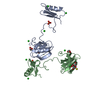
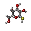
 Type: D-saccharide, beta linking / Mass: 196.221 Da / Num. of mol.: 2 / Source method: obtained synthetically / Formula: C6H12O5S


 Mass: 35.453 Da / Num. of mol.: 9 / Source method: obtained synthetically / Formula: Cl
 Mass: 96.063 Da / Num. of mol.: 2 / Source method: obtained synthetically / Formula: SO4
 Mass: 92.094 Da / Num. of mol.: 2 / Source method: obtained synthetically / Formula: C3H8O3
 Sample preparation
 Processing