- PDB-2yii: Manipulating the regioselectivity of phenylalanine aminomutase: n... -
+
Open data
ID or keywords:
Loading...
-
Basic information
Entry
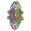
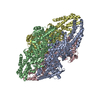
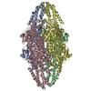
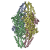
Database: PDB / ID: 2yii
Title
Manipulating the regioselectivity of phenylalanine aminomutase: new insights into the reaction mechanism of MIO-dependent enzymes from structure-guided directed evolution
Components
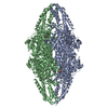
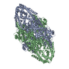
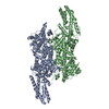
PHENYLALANINE AMMONIA-LYASE
Keywords
LYASE
Function / homology
Function and homology information
phenylalanine aminomutase (L-beta-phenylalanine-forming) / intramolecular aminotransferase activity / paclitaxel biosynthetic process / phenylalanine ammonia-lyase / : / cinnamic acid biosynthetic process / phenylalanine ammonia-lyase activity / alkaloid biosynthetic process / L-phenylalanine catabolic process / protein homotetramerization / cytoplasm Similarity search - Function
Mass: 77553.633 Da / Num. of mol.: 4 Source method: isolated from a genetically manipulated source Details: THIOESTER BOND BETWEEN THE MDO PROSTHETIC GROUP AND A MOLECULE OF BETA-MERCAPTOETHANOL, DISULFIDE BRIDGE BETWEEN RESIDUE C89 AND A BETA-MERCAPTOETHANOL MOLECULE, COVALENT BOND BETWEEN MDO 175 AND RESIDUE D178 Source: (gene. exp.) TAXUS WALLICHIANA VAR. CHINENSIS (Chinese yew) Plasmid: PBAD / Production host: ESCHERICHIA COLI (E. coli) / Strain (production host): K-12 / Variant (production host): TOP10 References: UniProt: Q68G84, EC: 5.4.3.-, phenylalanine ammonia-lyase
Resolution: 2.18→98.31 Å / Cor.coef. Fo:Fc: 0.954 / Cor.coef. Fo:Fc free: 0.932 / SU B: 5.134 / SU ML: 0.132 / Cross valid method: THROUGHOUT / ESU R: 0.254 / ESU R Free: 0.188 / Stereochemistry target values: MAXIMUM LIKELIHOOD / Details: HYDROGENS HAVE BEEN ADDED IN THE RIDING POSITIONS.
Rfactor
Num. reflection
% reflection
Selection details
Rfree
0.2145
7140
5 %
RANDOM
Rwork
0.17875
-
-
-
obs
0.18053
135680
97.89 %
-
Solvent computation
Ion probe radii: 0.8 Å / Shrinkage radii: 0.8 Å / VDW probe radii: 1.4 Å / Solvent model: BABINET MODEL WITH MASK
 Movie
Movie Controller

 Yorodumi
 Open data
 Basic information
 Components
 Keywords
 Function and homology information
 TAXUS WALLICHIANA VAR. CHINENSIS (Chinese yew)
 X-RAY DIFFRACTION /
  Authors
 Citation
 Structure visualization
 Downloads & links
 Other downloads


 PDBj
PDBj


 Assembly
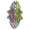


 Mass: 78.133 Da / Num. of mol.: 8 / Source method: obtained synthetically / Formula: C2H6OS
 Mass: 46.025 Da / Num. of mol.: 4 / Source method: obtained synthetically / Formula: CH2O2
 Mass: 92.094 Da / Num. of mol.: 1 / Source method: obtained synthetically / Formula: C3H8O3
 Mass: 18.015 Da / Num. of mol.: 834 / Source method: isolated from a natural source / Formula: H2O
 Sample preparation
 / Beamline: ID14-1 / Wavelength: 0.9334
  Processing