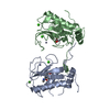
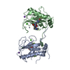
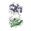
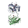
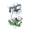
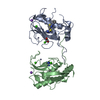
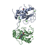
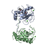
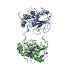
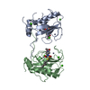
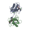
[English] 日本語
Yorodumi
- PDB-2yig: MMP13 in complex with a novel selective non zinc binding inhibitor -

+
Open data


ID or keywords:

Loading...

-
Basic information

Entry
Database: PDB / ID: 2yig
TitleMMP13 in complex with a novel selective non zinc binding inhibitor
ComponentsCOLLAGENASE 3
KeywordsHYDROLASE / COLLAGENASE 3 / MMP-13 / MATRIXMETALLOPROTEASE
Function / homology
Function and homology information


growth plate cartilage development / RUNX2 regulates genes involved in cell migration / endochondral ossification / Hydrolases; Acting on peptide bonds (peptidases); Metalloendopeptidases / bone morphogenesis / Assembly of collagen fibrils and other multimeric structures / bone mineralization / Activation of Matrix Metalloproteinases / response to amyloid-beta / Collagen degradation ...growth plate cartilage development / RUNX2 regulates genes involved in cell migration / endochondral ossification / Hydrolases; Acting on peptide bonds (peptidases); Metalloendopeptidases / bone morphogenesis / Assembly of collagen fibrils and other multimeric structures / bone mineralization / Activation of Matrix Metalloproteinases / response to amyloid-beta / Collagen degradation / collagen catabolic process / extracellular matrix disassembly / collagen binding / Degradation of the extracellular matrix / extracellular matrix organization / extracellular matrix / metalloendopeptidase activity / endopeptidase activity / serine-type endopeptidase activity / calcium ion binding / proteolysis / extracellular space / zinc ion binding / extracellular region
Similarity search - Function
Peptidoglycan binding-like / Hemopexin, conserved site / Hemopexin domain signature. / Hemopexin-like domain / Peptidase M10A, cysteine switch, zinc binding site / Matrixins cysteine switch. / Putative peptidoglycan binding domain / Hemopexin-like repeats / Hemopexin-like domain superfamily / Hemopexin ...Peptidoglycan binding-like / Hemopexin, conserved site / Hemopexin domain signature. / Hemopexin-like domain / Peptidase M10A, cysteine switch, zinc binding site / Matrixins cysteine switch. / Putative peptidoglycan binding domain / Hemopexin-like repeats / Hemopexin-like domain superfamily / Hemopexin / Hemopexin repeat profile. / Hemopexin-like repeats. / Peptidase M10A / Peptidase M10A, catalytic domain / Peptidase M10, metallopeptidase / Matrixin / PGBD-like superfamily / Peptidase, metallopeptidase / Zinc-dependent metalloprotease / Collagenase (Catalytic Domain) / Collagenase (Catalytic Domain) / Metallopeptidase, catalytic domain superfamily / Neutral zinc metallopeptidases, zinc-binding region signature. / 3-Layer(aba) Sandwich / Alpha Beta
Similarity search - Domain/homology
Chem-5EL / Collagenase 3
Similarity search - Component
Biological speciesHOMO SAPIENS (human)
MethodX-RAY DIFFRACTION / SYNCHROTRON / MOLECULAR REPLACEMENT / Resolution: 1.7 Å
AuthorsGerhardt, S. / Hargreaves, D.
CitationJournal: Bioorg.Med.Chem.Lett. / Year: 2011
Title: Selective Non Zinc Binding Inhibitors of Mmp13.
Authors: Savi, C.D. / Morley, A.D. / Ting, A. / Nash, I. / Karabelas, K. / Wood, C.M. / James, M. / Norris, S.J. / Karoutchi, G. / Rankine, N. / Hamlin, G. / Macfaul, P.A. / Ryan, D. / Baker, S.V. / ...Authors: Savi, C.D. / Morley, A.D. / Ting, A. / Nash, I. / Karabelas, K. / Wood, C.M. / James, M. / Norris, S.J. / Karoutchi, G. / Rankine, N. / Hamlin, G. / Macfaul, P.A. / Ryan, D. / Baker, S.V. / Hargreaves, D. / Gerhardt, S.
History
DepositionMay 13, 2011Deposition site: PDBE / Processing site: PDBE
Revision 1.0Jun 29, 2011Provider: repository / Type: Initial release
Revision 1.1Jul 7, 2011Group: Version format compliance
Revision 1.2Jul 13, 2011Group: Version format compliance
Revision 1.3Jul 12, 2017Group: Derived calculations / Category: pdbx_struct_conn_angle / struct_conn
Item: _pdbx_struct_conn_angle.ptnr1_auth_asym_id / _pdbx_struct_conn_angle.ptnr1_auth_comp_id ..._pdbx_struct_conn_angle.ptnr1_auth_asym_id / _pdbx_struct_conn_angle.ptnr1_auth_comp_id / _pdbx_struct_conn_angle.ptnr1_auth_seq_id / _pdbx_struct_conn_angle.ptnr1_label_asym_id / _pdbx_struct_conn_angle.ptnr1_label_atom_id / _pdbx_struct_conn_angle.ptnr1_label_comp_id / _pdbx_struct_conn_angle.ptnr1_label_seq_id / _pdbx_struct_conn_angle.ptnr1_symmetry / _pdbx_struct_conn_angle.ptnr2_auth_comp_id / _pdbx_struct_conn_angle.ptnr2_auth_seq_id / _pdbx_struct_conn_angle.ptnr2_label_asym_id / _pdbx_struct_conn_angle.ptnr2_label_atom_id / _pdbx_struct_conn_angle.ptnr2_label_comp_id / _pdbx_struct_conn_angle.ptnr3_auth_asym_id / _pdbx_struct_conn_angle.ptnr3_auth_comp_id / _pdbx_struct_conn_angle.ptnr3_auth_seq_id / _pdbx_struct_conn_angle.ptnr3_label_asym_id / _pdbx_struct_conn_angle.ptnr3_label_atom_id / _pdbx_struct_conn_angle.ptnr3_label_comp_id / _pdbx_struct_conn_angle.ptnr3_label_seq_id / _pdbx_struct_conn_angle.ptnr3_symmetry / _pdbx_struct_conn_angle.value / _struct_conn.pdbx_dist_value / _struct_conn.ptnr1_auth_asym_id / _struct_conn.ptnr1_auth_comp_id / _struct_conn.ptnr1_auth_seq_id / _struct_conn.ptnr1_label_asym_id / _struct_conn.ptnr1_label_atom_id / _struct_conn.ptnr1_label_comp_id / _struct_conn.ptnr1_label_seq_id / _struct_conn.ptnr2_auth_asym_id / _struct_conn.ptnr2_auth_comp_id / _struct_conn.ptnr2_auth_seq_id / _struct_conn.ptnr2_label_asym_id / _struct_conn.ptnr2_label_atom_id / _struct_conn.ptnr2_label_comp_id / _struct_conn.ptnr2_label_seq_id / _struct_conn.ptnr2_symmetry
Revision 1.4May 8, 2024Group: Data collection / Database references ...Data collection / Database references / Derived calculations / Other
Category: chem_comp_atom / chem_comp_bond ...chem_comp_atom / chem_comp_bond / database_2 / pdbx_database_status / pdbx_struct_conn_angle / struct_conn
Item: _database_2.pdbx_DOI / _database_2.pdbx_database_accession ..._database_2.pdbx_DOI / _database_2.pdbx_database_accession / _pdbx_database_status.status_code_sf / _pdbx_struct_conn_angle.ptnr1_auth_asym_id / _pdbx_struct_conn_angle.ptnr1_auth_comp_id / _pdbx_struct_conn_angle.ptnr1_auth_seq_id / _pdbx_struct_conn_angle.ptnr1_label_asym_id / _pdbx_struct_conn_angle.ptnr1_label_atom_id / _pdbx_struct_conn_angle.ptnr1_label_comp_id / _pdbx_struct_conn_angle.ptnr1_label_seq_id / _pdbx_struct_conn_angle.ptnr1_symmetry / _pdbx_struct_conn_angle.ptnr2_auth_asym_id / _pdbx_struct_conn_angle.ptnr2_auth_comp_id / _pdbx_struct_conn_angle.ptnr2_auth_seq_id / _pdbx_struct_conn_angle.ptnr2_label_asym_id / _pdbx_struct_conn_angle.ptnr2_label_atom_id / _pdbx_struct_conn_angle.ptnr2_label_comp_id / _pdbx_struct_conn_angle.ptnr3_auth_asym_id / _pdbx_struct_conn_angle.ptnr3_auth_comp_id / _pdbx_struct_conn_angle.ptnr3_auth_seq_id / _pdbx_struct_conn_angle.ptnr3_label_asym_id / _pdbx_struct_conn_angle.ptnr3_label_atom_id / _pdbx_struct_conn_angle.ptnr3_label_comp_id / _pdbx_struct_conn_angle.ptnr3_label_seq_id / _pdbx_struct_conn_angle.ptnr3_symmetry / _pdbx_struct_conn_angle.value / _struct_conn.pdbx_dist_value / _struct_conn.ptnr1_auth_asym_id / _struct_conn.ptnr1_auth_comp_id / _struct_conn.ptnr1_auth_seq_id / _struct_conn.ptnr1_label_asym_id / _struct_conn.ptnr1_label_atom_id / _struct_conn.ptnr1_label_comp_id / _struct_conn.ptnr1_label_seq_id / _struct_conn.ptnr1_symmetry / _struct_conn.ptnr2_auth_asym_id / _struct_conn.ptnr2_auth_comp_id / _struct_conn.ptnr2_auth_seq_id / _struct_conn.ptnr2_label_asym_id / _struct_conn.ptnr2_label_atom_id / _struct_conn.ptnr2_label_comp_id / _struct_conn.ptnr2_symmetry

-
Structure visualization

Structure viewerMolecule:
MolmilJmol/JSmol

Downloads & links

-
Assembly

Deposited unit
A: COLLAGENASE 3
B: COLLAGENASE 3
hetero molecules


Theoretical massNumber of molelcules
Total (without water)39,86915
Polymers38,4712
Non-polymers1,39813
Water5,350297
1


  • Idetical with deposited unit
  • defined by author&software
TypeNameSymmetry operationNumber
identity operation1_555x,y,z1
Buried area3250 Å2
ΔGint-148.2 kcal/mol
Surface area16040 Å2
MethodPISA
Unit cell
Length a, b, c (Å)134.417, 36.074, 95.206
Angle α, β, γ (deg.)90.00, 131.49, 90.00
Int Tables number5
Space group name H-MC121

-
Components

-
Protein , 1 types, 2 molecules AB

#1: Protein COLLAGENASE 3 / MATRIX METALLOPROTEINASE-13 / MMP-13


Mass: 19235.383 Da / Num. of mol.: 2 / Fragment: CATALYTIC DOMAIN, RESIDUES 104-274
Source method: isolated from a genetically manipulated source
Source: (gene. exp.) HOMO SAPIENS (human) / Plasmid: PT7#3.3 / Production host: ESCHERICHIA COLI (E. coli) / Strain (production host): BL21 / Variant (production host): PLYSS
References: UniProt: P45452, Hydrolases; Acting on peptide bonds (peptidases); Metalloendopeptidases

-
Non-polymers , 5 types, 310 molecules

#2: Chemical ChemComp-5EL / 4-(4-{[(3S)-3-HYDROXY-1-AZABICYCLO[2.2.2]OCT-3-YL]ETHYNYL}PHENOXY)-N-(PYRIDIN-4-YLMETHYL)BENZAMIDE


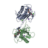
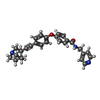
Mass: 453.532 Da / Num. of mol.: 2 / Source method: obtained synthetically / Formula: C28H27N3O3
#3: Chemical
ChemComp-ZN / ZINC ION


Mass: 65.409 Da / Num. of mol.: 4 / Source method: obtained synthetically / Formula: Zn
#4: Chemical
ChemComp-CA / CALCIUM ION


Mass: 40.078 Da / Num. of mol.: 4 / Source method: obtained synthetically / Formula: Ca
#5: Chemical ChemComp-NA / SODIUM ION


Mass: 22.990 Da / Num. of mol.: 3 / Source method: obtained synthetically / Formula: Na
#6: Water ChemComp-HOH / water


Mass: 18.015 Da / Num. of mol.: 297 / Source method: isolated from a natural source / Formula: H2O

-
Experimental details

-
Experiment

ExperimentMethod: X-RAY DIFFRACTION / Number of used crystals: 1

-
Sample preparation

CrystalDensity Matthews: 2.26 Å3/Da / Density % sol: 45.61 % / Description: NONE
Crystal growDetails: 10-27% PEG 3350, 1.5 M AMMONIUM FORMATE, 0.1 M TRIS/HCL PH 8.5

-
Data collection

DiffractionMean temperature: 100 K
Diffraction sourceSource: SYNCHROTRON / Site: ESRF / Beamline: BM14 / Wavelength: 1.0332
DetectorType: MARMOSAIC 225 mm CCD / Detector: CCD / Date: Mar 2, 2007
RadiationProtocol: SINGLE WAVELENGTH / Monochromatic (M) / Laue (L): M / Scattering type: x-ray
Radiation wavelengthWavelength: 1.0332 Å / Relative weight: 1
ReflectionResolution: 1.7→67.12 Å / Num. obs: 36370 / % possible obs: 95.9 % / Observed criterion σ(I): 0 / Redundancy: 1.9 % / Rmerge(I) obs: 0.07 / Net I/σ(I): 9.4
Reflection shellResolution: 1.7→1.74 Å / Redundancy: 1.9 % / Rmerge(I) obs: 0.35 / Mean I/σ(I) obs: 2.1 / % possible all: 95.9

-
Processing

Software
NameVersionClassification
REFMAC5.6.0113refinement
MOSFLMdata reduction
SCALAdata scaling
AMoREphasing
RefinementMethod to determine structure: MOLECULAR REPLACEMENT / Resolution: 1.7→67.12 Å / Cor.coef. Fo:Fc: 0.953 / Cor.coef. Fo:Fc free: 0.927 / SU B: 4.007 / SU ML: 0.075 / Cross valid method: THROUGHOUT / ESU R: 0.12 / ESU R Free: 0.115 / Stereochemistry target values: MAXIMUM LIKELIHOOD
Details: HYDROGENS HAVE BEEN ADDED IN THE RIDING POSITIONS. RESIDUES FROM SER A 250 TO PHE A 252 ARE DISORDERED. NCS TYPE: LOCAL, GROUP A 104-268, B 104-268, COUNT 211, RMS 0.15, WEIGHT 0.05.
RfactorNum. reflection% reflectionSelection details
Rfree0.22055 1830 5 %RANDOM
Rwork0.18441 ---
obs0.18624 34539 95.35 %-
Solvent computationIon probe radii: 0.8 Å / Shrinkage radii: 0.8 Å / VDW probe radii: 1.2 Å / Solvent model: MASK
Displacement parametersBiso mean: 15.38 Å2
Baniso -1Baniso -2Baniso -3
1-0.9 Å20 Å20.27 Å2
2---0.63 Å20 Å2
3---0.08 Å2
Refinement stepCycle: LAST / Resolution: 1.7→67.12 Å
ProteinNucleic acidLigandSolventTotal
Num. atoms2648 0 79 297 3024
Refine LS restraints
Refine-IDTypeDev idealDev ideal targetNumber
X-RAY DIFFRACTIONr_bond_refined_d0.0210.022818
X-RAY DIFFRACTIONr_bond_other_d
X-RAY DIFFRACTIONr_angle_refined_deg1.8351.9673833
X-RAY DIFFRACTIONr_angle_other_deg
X-RAY DIFFRACTIONr_dihedral_angle_1_deg6.4485330
X-RAY DIFFRACTIONr_dihedral_angle_2_deg36.25124.015132
X-RAY DIFFRACTIONr_dihedral_angle_3_deg14.22615403
X-RAY DIFFRACTIONr_dihedral_angle_4_deg21.66156
X-RAY DIFFRACTIONr_chiral_restr0.1470.2377
X-RAY DIFFRACTIONr_gen_planes_refined0.0120.0212306
X-RAY DIFFRACTIONr_gen_planes_other
X-RAY DIFFRACTIONr_nbd_refined
X-RAY DIFFRACTIONr_nbd_other
X-RAY DIFFRACTIONr_nbtor_refined
X-RAY DIFFRACTIONr_nbtor_other
X-RAY DIFFRACTIONr_xyhbond_nbd_refined
X-RAY DIFFRACTIONr_xyhbond_nbd_other
X-RAY DIFFRACTIONr_metal_ion_refined
X-RAY DIFFRACTIONr_metal_ion_other
X-RAY DIFFRACTIONr_symmetry_vdw_refined
X-RAY DIFFRACTIONr_symmetry_vdw_other
X-RAY DIFFRACTIONr_symmetry_hbond_refined
X-RAY DIFFRACTIONr_symmetry_hbond_other
X-RAY DIFFRACTIONr_symmetry_metal_ion_refined
X-RAY DIFFRACTIONr_symmetry_metal_ion_other
X-RAY DIFFRACTIONr_mcbond_it
X-RAY DIFFRACTIONr_mcbond_other
X-RAY DIFFRACTIONr_mcangle_it
X-RAY DIFFRACTIONr_mcangle_other
X-RAY DIFFRACTIONr_scbond_it
X-RAY DIFFRACTIONr_scbond_other
X-RAY DIFFRACTIONr_scangle_it
X-RAY DIFFRACTIONr_scangle_other
X-RAY DIFFRACTIONr_long_range_B_refined
X-RAY DIFFRACTIONr_long_range_B_other
X-RAY DIFFRACTIONr_rigid_bond_restr
X-RAY DIFFRACTIONr_sphericity_free
X-RAY DIFFRACTIONr_sphericity_bonded
LS refinement shellResolution: 1.7→1.744 Å / Total num. of bins used: 20
RfactorNum. reflection% reflection
Rfree0.359 122 -
Rwork0.281 2415 -
obs--95.38 %
Refinement TLS params.

Method: refined / Refine-ID: X-RAY DIFFRACTION

IDL112)L122)L132)L222)L232)L332)S11 (Å °)S12 (Å °)S13 (Å °)S21 (Å °)S22 (Å °)S23 (Å °)S31 (Å °)S32 (Å °)S33 (Å °)T112)T122)T132)T222)T232)T332)Origin x (Å)Origin y (Å)Origin z (Å)
10.96110.0753-0.74080.80880.08881.6235-0.01490.0375-0.0079-0.08660.0255-0.00820.0376-0.0011-0.01060.01820.0025-0.0060.0055-0.00480.005925.8613.820615.7458
20.7895-0.15930.04550.7171-0.03091.21880.03640.0554-0.0362-0.1493-0.0058-0.02650.04970.0508-0.03060.035-0.0016-0.00110.0185-0.00110.035855.9048-4.016825.9177
Refinement TLS group
IDRefine-IDRefine TLS-IDAuth asym-IDAuth seq-ID
1X-RAY DIFFRACTION1A104 - 272
2X-RAY DIFFRACTION2B104 - 269

+
About Yorodumi

-
News

-
Feb 9, 2022. New format data for meta-information of EMDB entries

New format data for meta-information of EMDB entries

  • Version 3 of the EMDB header file is now the official format.
  • The previous official version 1.9 will be removed from the archive.

Related info.:EMDB header

External links:wwPDB to switch to version 3 of the EMDB data model

-
Aug 12, 2020. Covid-19 info

Covid-19 info

URL: https://pdbjlvh1.pdbj.org/emnavi/covid19.php

New page: Covid-19 featured information page in EM Navigator.

Related info.:Covid-19 info / Mar 5, 2020. Novel coronavirus structure data

+
Mar 5, 2020. Novel coronavirus structure data

Novel coronavirus structure data

Related info.:Yorodumi Speices / Aug 12, 2020. Covid-19 info

External links:COVID-19 featured content - PDBj / Molecule of the Month (242):Coronavirus Proteases

+
Jan 31, 2019. EMDB accession codes are about to change! (news from PDBe EMDB page)

EMDB accession codes are about to change! (news from PDBe EMDB page)

  • The allocation of 4 digits for EMDB accession codes will soon come to an end. Whilst these codes will remain in use, new EMDB accession codes will include an additional digit and will expand incrementally as the available range of codes is exhausted. The current 4-digit format prefixed with “EMD-” (i.e. EMD-XXXX) will advance to a 5-digit format (i.e. EMD-XXXXX), and so on. It is currently estimated that the 4-digit codes will be depleted around Spring 2019, at which point the 5-digit format will come into force.
  • The EM Navigator/Yorodumi systems omit the EMD- prefix.

Related info.:Q: What is EMD? / ID/Accession-code notation in Yorodumi/EM Navigator

External links:EMDB Accession Codes are Changing Soon! / Contact to PDBj

+
Jul 12, 2017. Major update of PDB

Major update of PDB

  • wwPDB released updated PDB data conforming to the new PDBx/mmCIF dictionary.
  • This is a major update changing the version number from 4 to 5, and with Remediation, in which all the entries are updated.
  • In this update, many items about electron microscopy experimental information are reorganized (e.g. em_software).
  • Now, EM Navigator and Yorodumi are based on the updated data.

External links:wwPDB Remediation / Enriched Model Files Conforming to OneDep Data Standards Now Available in the PDB FTP Archive

-
Yorodumi

Thousand views of thousand structures

  • Yorodumi is a browser for structure data from EMDB, PDB, SASBDB, etc.
  • This page is also the successor to EM Navigator detail page, and also detail information page/front-end page for Omokage search.
  • The word "yorodu" (or yorozu) is an old Japanese word meaning "ten thousand". "mi" (miru) is to see.

Related info.:EMDB / PDB / SASBDB / Comparison of 3 databanks / Yorodumi Search / Aug 31, 2016. New EM Navigator & Yorodumi / Yorodumi Papers / Jmol/JSmol / Function and homology information / Changes in new EM Navigator and Yorodumi

Read more