Type: D-saccharide / Mass: 240.274 Da / Num. of mol.: 2 / Source method: obtained synthetically / Formula: C8H16O6S
Compound details
ENGINEERED RESIDUE IN CHAIN A, CYS 117 TO SER ENGINEERED RESIDUE IN CHAIN A, ALA 122 TO CYS ...ENGINEERED RESIDUE IN CHAIN A, CYS 117 TO SER ENGINEERED RESIDUE IN CHAIN A, ALA 122 TO CYS ENGINEERED RESIDUE IN CHAIN A, CYS 148 TO ALA ENGINEERED RESIDUE IN CHAIN A, CYS 154 TO VAL ENGINEERED RESIDUE IN CHAIN A, CYS 176 TO MET ENGINEERED RESIDUE IN CHAIN A, CYS 234 TO SER ENGINEERED RESIDUE IN CHAIN A, CYS 333 TO SER ENGINEERED RESIDUE IN CHAIN A, CYS 353 TO ALA ENGINEERED RESIDUE IN CHAIN A, CYS 355 TO ALA ENGINEERED RESIDUE IN CHAIN B, CYS 117 TO SER ENGINEERED RESIDUE IN CHAIN B, ALA 122 TO CYS ENGINEERED RESIDUE IN CHAIN B, CYS 148 TO ALA ENGINEERED RESIDUE IN CHAIN B, CYS 154 TO VAL ENGINEERED RESIDUE IN CHAIN B, CYS 176 TO MET ENGINEERED RESIDUE IN CHAIN B, CYS 234 TO SER ENGINEERED RESIDUE IN CHAIN B, CYS 333 TO SER ENGINEERED RESIDUE IN CHAIN B, CYS 353 TO ALA ENGINEERED RESIDUE IN CHAIN B, CYS 355 TO ALA
Has protein modification
Y
-
Experimental details
-
Experiment
Experiment
Method: X-RAY DIFFRACTION / Number of used crystals: 3
-
Sample preparation
Crystal
Density Matthews: 7.16 Å3/Da / Density % sol: 82.69 % Description: THE DATA DISPLAYED STRONG ANISOTROPY. THE NON- CORRECTED DATA WAS DEPOSITED IN THE PDB, WHILE THE CORRECTED DATA WAS INITIALLY USED TO SOLVE THE STRUCTURE, WITH BETTER DATA STATISTICS IN ...Description: THE DATA DISPLAYED STRONG ANISOTROPY. THE NON- CORRECTED DATA WAS DEPOSITED IN THE PDB, WHILE THE CORRECTED DATA WAS INITIALLY USED TO SOLVE THE STRUCTURE, WITH BETTER DATA STATISTICS IN THE HIGH RESOLUTION SHELLS.
In the structure databanks used in Yorodumi, some data are registered as the other names, "COVID-19 virus" and "2019-nCoV". Here are the details of the virus and the list of structure data.
Jan 31, 2019. EMDB accession codes are about to change! (news from PDBe EMDB page)
EMDB accession codes are about to change! (news from PDBe EMDB page)
The allocation of 4 digits for EMDB accession codes will soon come to an end. Whilst these codes will remain in use, new EMDB accession codes will include an additional digit and will expand incrementally as the available range of codes is exhausted. The current 4-digit format prefixed with “EMD-” (i.e. EMD-XXXX) will advance to a 5-digit format (i.e. EMD-XXXXX), and so on. It is currently estimated that the 4-digit codes will be depleted around Spring 2019, at which point the 5-digit format will come into force.
The EM Navigator/Yorodumi systems omit the EMD- prefix.
Related info.:Q: What is EMD? / ID/Accession-code notation in Yorodumi/EM Navigator
Yorodumi is a browser for structure data from EMDB, PDB, SASBDB, etc.
This page is also the successor to EM Navigator detail page, and also detail information page/front-end page for Omokage search.
The word "yorodu" (or yorozu) is an old Japanese word meaning "ten thousand". "mi" (miru) is to see.
Related info.:EMDB / PDB / SASBDB / Comparison of 3 databanks / Yorodumi Search / Aug 31, 2016. New EM Navigator & Yorodumi / Yorodumi Papers / Jmol/JSmol / Function and homology information / Changes in new EM Navigator and Yorodumi
 Movie
Movie Controller

 Yorodumi
 Open data
 Basic information
 Components
 Keywords
 Function and homology information
 X-RAY DIFFRACTION /
  Authors
 Citation
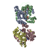
 Structure visualization
 Downloads & links
 Other downloads


 PDBj
PDBj


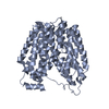
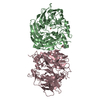
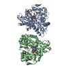
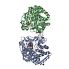
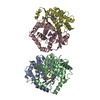
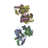
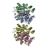
 Assembly
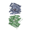

 Mass: 137.327 Da / Num. of mol.: 2 / Source method: obtained synthetically / Formula: Ba
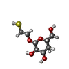
 Type: D-saccharide / Mass: 240.274 Da / Num. of mol.: 2 / Source method: obtained synthetically / Formula: C8H16O6S
 Sample preparation
 / Beamline: 5.0.2 / Wavelength: 1
  Processing