 Movie
Movie Controller

[English] 日本語
 Yorodumi
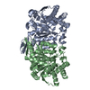
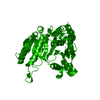
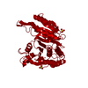
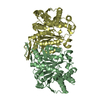
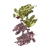
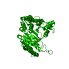
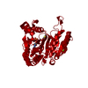
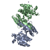
- PDB-2y3z: Structure of Isopropylmalate dehydrogenase from Thermus thermophi... -
+ Open data

- Basic information
| Entry | Database: PDB / ID: 2y3z | ||||||
|---|---|---|---|---|---|---|---|
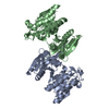
| Title | Structure of Isopropylmalate dehydrogenase from Thermus thermophilus - apo enzyme | ||||||
 Components Components | 3-ISOPROPYLMALATE DEHYDROGENASE | ||||||
 Keywords Keywords | OXIDOREDUCTASE / LEUB / LEUCINE BIOSYNTHESIS | ||||||
| Function / homology |  Function and homology information Function and homology information3-isopropylmalate dehydrogenase / 3-isopropylmalate dehydrogenase activity / L-leucine biosynthetic process / NAD binding / magnesium ion binding / identical protein binding / cytosol Similarity search - Function | ||||||
| Biological species |   THERMUS THERMOPHILUS (bacteria) THERMUS THERMOPHILUS (bacteria) | ||||||
| Method |  X-RAY DIFFRACTION / SYNCHROTRON / MOLECULAR REPLACEMENT / Resolution: 1.83 Å X-RAY DIFFRACTION / SYNCHROTRON / MOLECULAR REPLACEMENT / Resolution: 1.83 Å | ||||||
 Authors Authors | Graczer, E. / merlin, A. / Singh, R.K. / Manikandan, K. / Zavodsky, P. / Weiss, M.S. / Vas, M. | ||||||
 Citation Citation | Journal: Mol.Biosyst. / Year: 2011 Title: Atomic Level Description of the Domain Closure in a Dimeric Enzyme: Thermus Thermophilus 3-Isopropylmalate Dehydrogenase. Authors: Graczer, E. / Merli, A. / Singh, R.K. / Karuppasamy, M. / Zavodszky, P. / Weiss, M.S. / Vas, M. #1: Journal: Acta Crystallogr.,Sect.F / Year: 2010 Title: Crystallization and Preliminary X-Ray Diffraction Analysis of Various Enzyme-Substrate Complexes of Isopropylmalate Dehydrogenase from Thermus Thermophilus. Authors: Merli, A. / Manikandan, K. / Graczer, E. / Schuldt, L. / Singh, R.K. / Zavodszky, P. / Vas, M. / Weiss, M.S. | ||||||
| History |
|
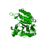
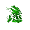
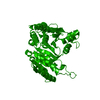
- Structure visualization
| Structure viewer | Molecule: MolmilJmol/JSmol |
|---|
- Downloads & links
-Download
| PDBx/mmCIF format | 2y3z.cif.gz | 153.1 KB | Display | PDBx/mmCIF format |
|---|---|---|---|---|
| PDB format | pdb2y3z.ent.gz | 120.6 KB | Display | PDB format |
| PDBx/mmJSON format | 2y3z.json.gz | Tree view | PDBx/mmJSON format | |
| Others |  Other downloads Other downloads |
-Validation report
| Arichive directory | https://data.pdbj.org/pub/pdb/validation_reports/y3/2y3zftp://data.pdbj.org/pub/pdb/validation_reports/y3/2y3z | HTTPS FTP |
|---|
-Related structure data
| Related structure data |  2y40C 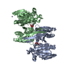 2y41C 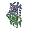 2y42C 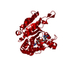 1hexS S: Starting model for refinement C: citing same article ( |
|---|---|
| Similar structure data |


-Links
 PDBj
PDBj

- Assembly
| Deposited unit | 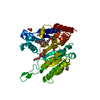
| ||||||||
|---|---|---|---|---|---|---|---|---|---|
| 1 | 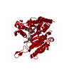
| ||||||||
| Unit cell |
|
-Components
| #1: Protein | Mass: 38398.883 Da / Num. of mol.: 1 Source method: isolated from a genetically manipulated source Details: ADDITIONAL AMINO ACIDS- N-TERMINAL MAS. C- TERMINAL AAALEHHHHHH Source: (gene. exp.) THERMUS THERMOPHILUS (bacteria) / Production host: References: UniProt: Q5SIY4, 3-isopropylmalate dehydrogenase | ||||||
|---|---|---|---|---|---|---|---|
| #2: Chemical | ChemComp-TRS /   Mass: 122.143 Da / Num. of mol.: 1 / Source method: obtained synthetically / Formula: C4H12NO3 / Comment: pH buffer*YM Mass: 122.143 Da / Num. of mol.: 1 / Source method: obtained synthetically / Formula: C4H12NO3 / Comment: pH buffer*YM | ||||||
| #3: Chemical |   Mass: 414.488 Da / Num. of mol.: 3 / Source method: obtained synthetically / Formula: C18H38O10 / Comment: precipitant*YM Mass: 414.488 Da / Num. of mol.: 3 / Source method: obtained synthetically / Formula: C18H38O10 / Comment: precipitant*YM#4: Chemical |   Mass: 92.094 Da / Num. of mol.: 3 / Source method: obtained synthetically / Formula: C3H8O3 Mass: 92.094 Da / Num. of mol.: 3 / Source method: obtained synthetically / Formula: C3H8O3#5: Water | ChemComp-HOH / |  Mass: 18.015 Da / Num. of mol.: 201 / Source method: isolated from a natural source / Formula: H2O Mass: 18.015 Da / Num. of mol.: 201 / Source method: isolated from a natural source / Formula: H2ONonpolymer details | POLYETHYLE | |
-Experimental details
-Experiment
| Experiment | Method: X-RAY DIFFRACTION / Number of used crystals: 1 |
|---|
- Sample preparation
| Crystal | Density Matthews: 3.62 Å3/Da / Density % sol: 66 % / Description: NONE |
|---|---|
| Crystal grow | pH: 9 / Details: 35% PEG-550-MME, 0.1 M TRIS-HCL PH 9.0, 0.1 M NACL |
-Data collection
| Diffraction | Mean temperature: 100 K |
|---|---|
| Diffraction source | Source: SYNCHROTRON / Site: ESRF  / Beamline: ID14-2 / Wavelength: 0.9334 / Beamline: ID14-2 / Wavelength: 0.9334 |
| Detector | Type: ADSC CCD / Detector: CCD |
| Radiation | Protocol: SINGLE WAVELENGTH / Monochromatic (M) / Laue (L): M / Scattering type: x-ray |
| Radiation wavelength | Wavelength: 0.9334 Å / Relative weight: 1 |
| Reflection | Resolution: 1.83→99 Å / Num. obs: 47324 / % possible obs: 99 % / Observed criterion σ(I): -3 / Redundancy: 6 % / Biso Wilson estimate: 29.1 Å2 / Rmerge(I) obs: 0.08 / Net I/σ(I): 16.7 |
| Reflection shell | Resolution: 1.83→1.86 Å / Redundancy: 4.5 % / Rmerge(I) obs: 0.65 / Mean I/σ(I) obs: 2.4 / % possible all: 91.2 |
- Processing
| Software |
| ||||||||||||||||||||||||||||||||||||||||||||||||||||||||||||||||||||||||||||||||||||||||||||||||||||||||||||||||||||||||||||||||||||||||||||||||||||||||||||||||||||||||||||||||||||||
|---|---|---|---|---|---|---|---|---|---|---|---|---|---|---|---|---|---|---|---|---|---|---|---|---|---|---|---|---|---|---|---|---|---|---|---|---|---|---|---|---|---|---|---|---|---|---|---|---|---|---|---|---|---|---|---|---|---|---|---|---|---|---|---|---|---|---|---|---|---|---|---|---|---|---|---|---|---|---|---|---|---|---|---|---|---|---|---|---|---|---|---|---|---|---|---|---|---|---|---|---|---|---|---|---|---|---|---|---|---|---|---|---|---|---|---|---|---|---|---|---|---|---|---|---|---|---|---|---|---|---|---|---|---|---|---|---|---|---|---|---|---|---|---|---|---|---|---|---|---|---|---|---|---|---|---|---|---|---|---|---|---|---|---|---|---|---|---|---|---|---|---|---|---|---|---|---|---|---|---|---|---|---|---|
| Refinement | Method to determine structure: MOLECULAR REPLACEMENT Starting model: PDB ENTRY 1HEX Resolution: 1.83→40 Å / Cor.coef. Fo:Fc: 0.969 / Cor.coef. Fo:Fc free: 0.956 / SU B: 4.53 / SU ML: 0.064 / Cross valid method: THROUGHOUT / ESU R: 0.09 / ESU R Free: 0.093 / Stereochemistry target values: MAXIMUM LIKELIHOOD / Details: HYDROGENS HAVE BEEN ADDED IN THE RIDING POSITIONS.
| ||||||||||||||||||||||||||||||||||||||||||||||||||||||||||||||||||||||||||||||||||||||||||||||||||||||||||||||||||||||||||||||||||||||||||||||||||||||||||||||||||||||||||||||||||||||
| Solvent computation | Ion probe radii: 0.8 Å / Shrinkage radii: 0.8 Å / VDW probe radii: 1.4 Å / Solvent model: BABINET MODEL WITH MASK | ||||||||||||||||||||||||||||||||||||||||||||||||||||||||||||||||||||||||||||||||||||||||||||||||||||||||||||||||||||||||||||||||||||||||||||||||||||||||||||||||||||||||||||||||||||||
| Displacement parameters | Biso mean: 32.74 Å2
| ||||||||||||||||||||||||||||||||||||||||||||||||||||||||||||||||||||||||||||||||||||||||||||||||||||||||||||||||||||||||||||||||||||||||||||||||||||||||||||||||||||||||||||||||||||||
| Refinement step | Cycle: LAST / Resolution: 1.83→40 Å
| ||||||||||||||||||||||||||||||||||||||||||||||||||||||||||||||||||||||||||||||||||||||||||||||||||||||||||||||||||||||||||||||||||||||||||||||||||||||||||||||||||||||||||||||||||||||
| Refine LS restraints |
|