 Movie
Movie Controller


+ Open data

- Basic information
| Entry | Database: PDB / ID: 1osi | ||||||
|---|---|---|---|---|---|---|---|
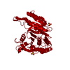
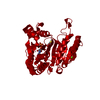
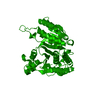
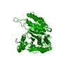
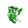
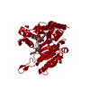
| Title | STRUCTURE OF 3-ISOPROPYLMALATE DEHYDROGENASE | ||||||
 Components Components | 3-ISOPROPYLMALATE DEHYDROGENASE | ||||||
 Keywords Keywords | OXIDOREDUCTASE / DEHYDROGENASE | ||||||
| Function / homology |  Function and homology information Function and homology information3-isopropylmalate dehydrogenase / 3-isopropylmalate dehydrogenase activity / L-leucine biosynthetic process / NAD binding / magnesium ion binding / identical protein binding / cytosol Similarity search - Function | ||||||
| Biological species |   Thermus thermophilus (bacteria) Thermus thermophilus (bacteria) | ||||||
| Method |  X-RAY DIFFRACTION / Resolution: 3 Å X-RAY DIFFRACTION / Resolution: 3 Å | ||||||
 Authors Authors | Qu, C. / Akanuma, S. / Moriyama, H. / Tanaka, N. / Oshima, T. | ||||||
 Citation Citation | Journal: Protein Eng. / Year: 1997 Title: A mutation at the interface between domains causes rearrangement of domains in 3-isopropylmalate dehydrogenase. Authors: Qu, C. / Akanuma, S. / Moriyama, H. / Tanaka, N. / Oshima, T. #1: Journal: J.Biochem.(Tokyo) / Year: 1990Title: Purification, Catalytic Properties, and Thermal Stability of Threo-Ds-3-Isopropylmalate Dehydrogenase Coded by Leub Gene from an Extreme Thermophile, Thermus Thermophilus Strain Hb8 Authors: Yamada, T. / Akutsu, N. / Miyazaki, K. / Kakinuma, K. / Yoshida, M. / Oshima, T. | ||||||
| History |
|
- Structure visualization
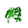
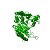
| Structure viewer | Molecule: MolmilJmol/JSmol |
|---|
- Downloads & links
-Download
| PDBx/mmCIF format | 1osi.cif.gz | 299.9 KB | Display | PDBx/mmCIF format |
|---|---|---|---|---|
| PDB format | pdb1osi.ent.gz | 247.8 KB | Display | PDB format |
| PDBx/mmJSON format | 1osi.json.gz | Tree view | PDBx/mmJSON format | |
| Others |  Other downloads Other downloads |
-Validation report
| Arichive directory | https://data.pdbj.org/pub/pdb/validation_reports/os/1osiftp://data.pdbj.org/pub/pdb/validation_reports/os/1osi | HTTPS FTP |
|---|
-Related structure data
-Links
 PDBj
PDBj

- Assembly
| Deposited unit | 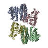
| ||||||||
|---|---|---|---|---|---|---|---|---|---|
| 1 | 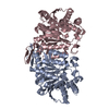
| ||||||||
| 2 | 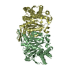
| ||||||||
| Unit cell |
| ||||||||
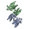
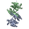
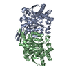
| Details | THE ASYMMETRIC UNIT CONTAINS TWO FUNCTIONAL DIMERS, FORMED BY THE SUBUNITS A AND C, B AND D. |
-Components
| #1: Protein | Mass: 36825.160 Da / Num. of mol.: 4 Source method: isolated from a genetically manipulated source Source: (gene. exp.) Thermus thermophilus (bacteria) / Strain: HB8 / Production host: unidentified (others)References: UniProt: Q5SIY4, 3-isopropylmalate dehydrogenase |
|---|
-Experimental details
-Experiment
| Experiment | Method: X-RAY DIFFRACTION / Number of used crystals: 1 |
|---|
- Sample preparation
| Crystal | Density Matthews: 2.72 Å3/Da / Density % sol: 58.2 % | |||||||||||||||||||||||||
|---|---|---|---|---|---|---|---|---|---|---|---|---|---|---|---|---|---|---|---|---|---|---|---|---|---|---|
| Crystal grow | pH: 4.8 Details: THE STRUCTURE SOLVED USING CRYSTALS GROWN FROM AMMONIUM SULFATE WAS DEPOSITED AS 1IPD AT PDB. THE CRYSTALLIZATION CONDITION IN THIS STRUCTURE WAS SIMILAR AS THE MUTANT A172L., pH 4.8 | |||||||||||||||||||||||||
| Crystal grow | *PLUS pH: 7.6 / Method: unknown | |||||||||||||||||||||||||
| Components of the solutions | *PLUS
|
-Data collection
| Diffraction | Mean temperature: 293 K |
|---|---|
| Diffraction source | Wavelength: 1.5418 |
| Detector | Type: RIGAKU RAXIS IIC / Detector: IMAGE PLATE / Date: Jul 15, 1995 |
| Radiation | Monochromatic (M) / Laue (L): M / Scattering type: x-ray |
| Radiation wavelength | Wavelength: 1.5418 Å / Relative weight: 1 |
| Reflection | Num. obs: 27930 / % possible obs: 93.8 % / Observed criterion σ(I): 1 / Redundancy: 0.927 % / Rmerge(I) obs: 0.054 |
- Processing
| Software |
| ||||||||||||||||||||||||||||||||||||||||||||||||||||||||||||
|---|---|---|---|---|---|---|---|---|---|---|---|---|---|---|---|---|---|---|---|---|---|---|---|---|---|---|---|---|---|---|---|---|---|---|---|---|---|---|---|---|---|---|---|---|---|---|---|---|---|---|---|---|---|---|---|---|---|---|---|---|---|
| Refinement | Resolution: 3→6 Å / σ(F): 2 /
| ||||||||||||||||||||||||||||||||||||||||||||||||||||||||||||
| Displacement parameters | Biso mean: 26.2 Å2 | ||||||||||||||||||||||||||||||||||||||||||||||||||||||||||||
| Refinement step | Cycle: LAST / Resolution: 3→6 Å
| ||||||||||||||||||||||||||||||||||||||||||||||||||||||||||||
| Refine LS restraints |
|
