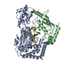
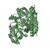
Journal: PLoS Pathog / Year: 2012 Title: The Vibrio cholerae colonization factor GbpA possesses a modular structure that governs binding to different host surfaces. Authors: Edmond Wong / Gustav Vaaje-Kolstad / Avishek Ghosh / Ramon Hurtado-Guerrero / Peter V Konarev / Adel F M Ibrahim / Dmitri I Svergun / Vincent G H Eijsink / Nabendu S Chatterjee / Daan M F van Aalten / Abstract: Vibrio cholerae is a bacterial pathogen that colonizes the chitinous exoskeleton of zooplankton as well as the human gastrointestinal tract. Colonization of these different niches involves an N- ...Vibrio cholerae is a bacterial pathogen that colonizes the chitinous exoskeleton of zooplankton as well as the human gastrointestinal tract. Colonization of these different niches involves an N-acetylglucosamine binding protein (GbpA) that has been reported to mediate bacterial attachment to both marine chitin and mammalian intestinal mucin through an unknown molecular mechanism. We report structural studies that reveal that GbpA possesses an unusual, elongated, four-domain structure, with domains 1 and 4 showing structural homology to chitin binding domains. A glycan screen revealed that GbpA binds to GlcNAc oligosaccharides. Structure-guided GbpA truncation mutants show that domains 1 and 4 of GbpA interact with chitin in vitro, whereas in vivo complementation studies reveal that domain 1 is also crucial for mucin binding and intestinal colonization. Bacterial binding studies show that domains 2 and 3 bind to the V. cholerae surface. Finally, mouse virulence assays show that only the first three domains of GbpA are required for colonization. These results explain how GbpA provides structural/functional modular interactions between V. cholerae, intestinal epithelium and chitinous exoskeletons.
In the structure databanks used in Yorodumi, some data are registered as the other names, "COVID-19 virus" and "2019-nCoV". Here are the details of the virus and the list of structure data.
Jan 31, 2019. EMDB accession codes are about to change! (news from PDBe EMDB page)
EMDB accession codes are about to change! (news from PDBe EMDB page)
The allocation of 4 digits for EMDB accession codes will soon come to an end. Whilst these codes will remain in use, new EMDB accession codes will include an additional digit and will expand incrementally as the available range of codes is exhausted. The current 4-digit format prefixed with “EMD-” (i.e. EMD-XXXX) will advance to a 5-digit format (i.e. EMD-XXXXX), and so on. It is currently estimated that the 4-digit codes will be depleted around Spring 2019, at which point the 5-digit format will come into force.
The EM Navigator/Yorodumi systems omit the EMD- prefix.
Related info.:Q: What is EMD? / ID/Accession-code notation in Yorodumi/EM Navigator
Yorodumi is a browser for structure data from EMDB, PDB, SASBDB, etc.
This page is also the successor to EM Navigator detail page, and also detail information page/front-end page for Omokage search.
The word "yorodu" (or yorozu) is an old Japanese word meaning "ten thousand". "mi" (miru) is to see.
Related info.:EMDB / PDB / SASBDB / Comparison of 3 databanks / Yorodumi Search / Aug 31, 2016. New EM Navigator & Yorodumi / Yorodumi Papers / Jmol/JSmol / Function and homology information / Changes in new EM Navigator and Yorodumi
 Movie
Movie Controller


 Open data
 Basic information
 Components
 Keywords
 Function and homology information
 VIBRIO CHOLERAE (bacteria)
 X-RAY DIFFRACTION /
  Authors
 Citation
 Structure visualization
 Downloads & links
 Other downloads
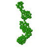


 PDBj
PDBj Assembly
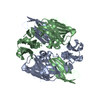
 Mass: 18.015 Da / Num. of mol.: 630 / Source method: isolated from a natural source / Formula: H2O
 Sample preparation
 / Beamline: BM14 / Wavelength: 0.933
  Processing