Mass: 483.156 Da / Num. of mol.: 2 / Source method: obtained synthetically / Formula: C9H16N3O14P3
-
Experimental details
-
Experiment
Experiment
Method: X-RAY DIFFRACTION / Number of used crystals: 1
-
Sample preparation
Crystal
Density Matthews: 2.51 Å3/Da / Density % sol: 51.04 % / Description: NONE
Crystal grow
Temperature: 293 K / Method: vapor diffusion, sitting drop Details: SITTING-DROP VAPOR-DIFFUSION METHOD AT 293 K. DROPS WITH 300 NANOLITERS PROTEIN SOLUTION (PROTEIN AT 10 MG/ML IN 20 MM BIS-TRIS-PROPANE, PH 6.5, 0.1 M NACL WITH 10 MM CTP AND 10 MM MGCL2) ...Details: SITTING-DROP VAPOR-DIFFUSION METHOD AT 293 K. DROPS WITH 300 NANOLITERS PROTEIN SOLUTION (PROTEIN AT 10 MG/ML IN 20 MM BIS-TRIS-PROPANE, PH 6.5, 0.1 M NACL WITH 10 MM CTP AND 10 MM MGCL2) AND 300 NANOLITERS SCREENING/RESERVOIR SOLUTION (10% (W/V) PEG 20, 000, 20% (V/V) MONOMETHYL ETHER PEG 550, 0.03 M CACL2, 0.03M MGCL2, AND 0.1 M BICINE/TRIZMA BASE, PH 8.5)
Protocol: SINGLE WAVELENGTH / Monochromatic (M) / Laue (L): M / Scattering type: x-ray
Radiation wavelength
Wavelength: 0.9334 Å / Relative weight: 1
Reflection
Resolution: 2.9→42 Å / Num. obs: 11201 / % possible obs: 99.2 % / Observed criterion σ(I): 0 / Redundancy: 7.1 % / Rmerge(I) obs: 0.13 / Net I/σ(I): 16.2
Reflection shell
Resolution: 2.9→2.98 Å / Redundancy: 7.3 % / Rmerge(I) obs: 0.49 / Mean I/σ(I) obs: 4 / % possible all: 98.9
-
Processing
Software
Name
Version
Classification
REFMAC
5.6.0060
refinement
XDS
datareduction
XSCALE
datascaling
PHASER
phasing
Refinement
Method to determine structure: MOLECULAR REPLACEMENT / Resolution: 2.9→41.83 Å / Cor.coef. Fo:Fc: 0.918 / Cor.coef. Fo:Fc free: 0.885 / SU B: 16.18 / SU ML: 0.314 / Cross valid method: THROUGHOUT / ESU R Free: 0.419 / Stereochemistry target values: MAXIMUM LIKELIHOOD / Details: HYDROGENS HAVE BEEN ADDED IN THE RIDING POSITIONS.
Rfactor
Num. reflection
% reflection
Selection details
Rfree
0.25337
560
5 %
RANDOM
Rwork
0.20833
-
-
-
obs
0.21065
10641
100 %
-
Solvent computation
Ion probe radii: 0.8 Å / Shrinkage radii: 0.8 Å / VDW probe radii: 1.2 Å / Solvent model: MASK
 Movie
Movie Controller

 Yorodumi
 Open data
 Basic information
 Components
 Keywords
 Function and homology information
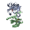
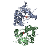
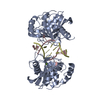
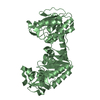
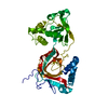
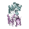
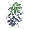
 MYCOBACTERIUM TUBERCULOSIS (bacteria)
 X-RAY DIFFRACTION /
  Authors
 Citation
 Structure visualization
 Downloads & links
 Other downloads
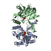


 PDBj
PDBj


 Assembly
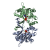

 Mass: 24.305 Da / Num. of mol.: 2 / Source method: obtained synthetically / Formula: Mg
 Mass: 483.156 Da / Num. of mol.: 2 / Source method: obtained synthetically / Formula: C9H16N3O14P3
 Sample preparation
 / Beamline: ID14-1 / Wavelength: 0.9334
  Processing