Mass: 18.015 Da / Num. of mol.: 274 / Source method: isolated from a natural source / Formula: H2O
-
Experimental details
-
Experiment
Experiment
Method: X-RAY DIFFRACTION / Number of used crystals: 1
-
Sample preparation
Crystal
Density Matthews: 2.04 Å3/Da / Density % sol: 39.83 % / Description: NONE
Crystal grow
Temperature: 293 K / Method: vapor diffusion, sitting drop Details: SITTING-DROP VAPOR-DIFFUSION METHOD AT 293 K. DROPS WITH 300 NANOLITERS PROTEIN SOLUTION (PROTEIN AT 40 MG/ML IN 20 MM BIS-TRIS-PROPANE, PH 6.5, 0.1 M NACL WITH 10 MM CMP, 10 MM MGCL2 AND 10 ...Details: SITTING-DROP VAPOR-DIFFUSION METHOD AT 293 K. DROPS WITH 300 NANOLITERS PROTEIN SOLUTION (PROTEIN AT 40 MG/ML IN 20 MM BIS-TRIS-PROPANE, PH 6.5, 0.1 M NACL WITH 10 MM CMP, 10 MM MGCL2 AND 10 MM ERYTHRITOL) AND 300 NANOLITERS SCREENING/RESERVOIR SOLUTION (10% (W/V) PEG 20,000, 20% (V/V) MONOMETHYL ETHER PEG 550, 0.03M NAF, 0.03M NAI, 0.03M NABR, AND 0.1M MOPS/NA-HEPES, PH 7.5)
 Movie
Movie Controller

 Yorodumi
 Open data
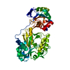
 Basic information
 Components
 Keywords
 Function and homology information
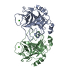
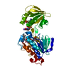
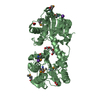
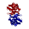
 MYCOBACTERIUM SMEGMATIS (bacteria)
 X-RAY DIFFRACTION /
  Authors
 Citation
 Structure visualization
 Downloads & links
 Other downloads
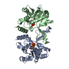


 PDBj
PDBj

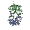
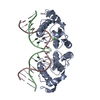
 Assembly

 Mass: 323.197 Da / Num. of mol.: 2 / Source method: obtained synthetically / Formula: C9H14N3O8P
 Mass: 18.015 Da / Num. of mol.: 274 / Source method: isolated from a natural source / Formula: H2O
 Sample preparation
 / Beamline: ID14-2 / Wavelength: 0.933
  Processing