 Movie
Movie Controller

[English] 日本語
 Yorodumi
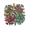
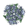
- PDB-1yac: THE 1.8 ANGSTROM CRYSTAL STRUCTURE OF THE YCAC GENE PRODUCT FROM ... -
+ Open data

- Basic information
| Entry | Database: PDB / ID: 1yac | ||||||
|---|---|---|---|---|---|---|---|
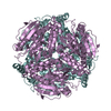
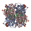
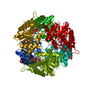
| Title | THE 1.8 ANGSTROM CRYSTAL STRUCTURE OF THE YCAC GENE PRODUCT FROM ESCHERICHIA COLI REVEALS AN OCTAMERIC HYDROLASE OF UNKNOWN SPECIFICITY | ||||||
 Components Components | YCAC GENE PRODUCT | ||||||
 Keywords Keywords | UNKNOWN BACTERIAL HYDROLASE / THREE LAYER ALPHA-BETA-ALPHA SANDWICH TOPOLOGY / ENTB HOMOLOG / CSHASE HOMOLOG | ||||||
| Function / homology |  Function and homology information Function and homology informationLyases / lyase activity / hydrolase activity / membrane / identical protein binding / cytosol Similarity search - Function | ||||||
| Biological species |  | ||||||
| Method |  X-RAY DIFFRACTION / SYNCHROTRON / MIR / Resolution: 1.8 Å X-RAY DIFFRACTION / SYNCHROTRON / MIR / Resolution: 1.8 Å | ||||||
 Authors Authors | Colovos, C. / Cascio, D. / Yeates, T.O. | ||||||
 Citation Citation | Journal: Structure / Year: 1998 Title: The 1.8 A crystal structure of the ycaC gene product from Escherichia coli reveals an octameric hydrolase of unknown specificity. Authors: Colovos, C. / Cascio, D. / Yeates, T.O. | ||||||
| History |
|
- Structure visualization
| Structure viewer | Molecule: MolmilJmol/JSmol |
|---|
- Downloads & links
-Download
| PDBx/mmCIF format | 1yac.cif.gz | 93.4 KB | Display | PDBx/mmCIF format |
|---|---|---|---|---|
| PDB format | pdb1yac.ent.gz | 72 KB | Display | PDB format |
| PDBx/mmJSON format | 1yac.json.gz | Tree view | PDBx/mmJSON format | |
| Others |  Other downloads Other downloads |
-Validation report
| Arichive directory | https://data.pdbj.org/pub/pdb/validation_reports/ya/1yacftp://data.pdbj.org/pub/pdb/validation_reports/ya/1yac | HTTPS FTP |
|---|
-Related structure data
| Similar structure data |
|---|

-Links
 PDBj
PDBj- Assembly
| Deposited unit | 
| |||||||||||||||
|---|---|---|---|---|---|---|---|---|---|---|---|---|---|---|---|---|
| 1 | 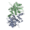
| |||||||||||||||
| Unit cell |
| |||||||||||||||
| Components on special symmetry positions |
| |||||||||||||||
| Noncrystallographic symmetry (NCS) | NCS oper: (Code: given Matrix: (-0.151263, 0.988474, -0.00626), Vector: |
-Components
| #1: Protein | Mass: 22978.121 Da / Num. of mol.: 2 Source method: isolated from a genetically manipulated source Source: (gene. exp.) #2: Water | ChemComp-HOH / |  Mass: 18.015 Da / Num. of mol.: 269 / Source method: isolated from a natural source / Formula: H2O Mass: 18.015 Da / Num. of mol.: 269 / Source method: isolated from a natural source / Formula: H2O |
|---|
-Experimental details
-Experiment
| Experiment | Method: X-RAY DIFFRACTION / Number of used crystals: 1 |
|---|
- Sample preparation
| Crystal | Density Matthews: 2.57 Å3/Da / Density % sol: 41 % | |||||||||||||||
|---|---|---|---|---|---|---|---|---|---|---|---|---|---|---|---|---|
| Crystal grow | *PLUS pH: 6 / Method: vapor diffusion, hanging drop | |||||||||||||||
| Components of the solutions | *PLUS
|
-Data collection
| Diffraction | Mean temperature: 90 K |
|---|---|
| Diffraction source | Source: SYNCHROTRON / Site: NSLS  / Beamline: X12B / Wavelength: 0.978 / Beamline: X12B / Wavelength: 0.978 |
| Detector | Type: ADSC / Detector: CCD / Date: Jul 9, 1997 / Details: MIRROR |
| Radiation | Monochromator: SI(111) / Monochromatic (M) / Laue (L): M / Scattering type: x-ray |
| Radiation wavelength | Wavelength: 0.978 Å / Relative weight: 1 |
| Reflection | Resolution: 1.8→40 Å / Num. obs: 45552 / % possible obs: 99.7 % / Observed criterion σ(I): 0 / Redundancy: 6.39 % / Biso Wilson estimate: 15.5 Å2 / Rmerge(I) obs: 0.076 / Net I/σ(I): 22.64 |
| Reflection shell | Resolution: 1.8→1.86 Å / Rmerge(I) obs: 0.26 / Mean I/σ(I) obs: 4.56 / % possible all: 97.6 |
| Reflection | *PLUS Num. obs: 45076 / % possible obs: 98.8 % / Num. measured all: 308775 / Rmerge(I) obs: 0.067 |
| Reflection shell | *PLUS % possible obs: 95.6 % / Mean I/σ(I) obs: 4.9 |
- Processing
| Software |
| ||||||||||||||||||||||||||||||||||||||||||||||||||||||||||||
|---|---|---|---|---|---|---|---|---|---|---|---|---|---|---|---|---|---|---|---|---|---|---|---|---|---|---|---|---|---|---|---|---|---|---|---|---|---|---|---|---|---|---|---|---|---|---|---|---|---|---|---|---|---|---|---|---|---|---|---|---|---|
| Refinement | Method to determine structure: MIR / Resolution: 1.8→8 Å / Data cutoff low absF: 0 / Cross valid method: THROUGHOUT / σ(F): 0
| ||||||||||||||||||||||||||||||||||||||||||||||||||||||||||||
| Displacement parameters | Biso mean: 14 Å2 | ||||||||||||||||||||||||||||||||||||||||||||||||||||||||||||
| Refinement step | Cycle: LAST / Resolution: 1.8→8 Å
| ||||||||||||||||||||||||||||||||||||||||||||||||||||||||||||
| Refine LS restraints |
| ||||||||||||||||||||||||||||||||||||||||||||||||||||||||||||
| Refine LS restraints NCS | NCS model details: RESTRAINTS | ||||||||||||||||||||||||||||||||||||||||||||||||||||||||||||
| LS refinement shell | Resolution: 1.8→1.88 Å / Total num. of bins used: 8
| ||||||||||||||||||||||||||||||||||||||||||||||||||||||||||||
| Xplor file |
| ||||||||||||||||||||||||||||||||||||||||||||||||||||||||||||
| Software | *PLUS Name: X-PLOR / Version: 3.843 / Classification: refinement | ||||||||||||||||||||||||||||||||||||||||||||||||||||||||||||
| Refinement | *PLUS Rfactor obs: 0.175 | ||||||||||||||||||||||||||||||||||||||||||||||||||||||||||||
| Solvent computation | *PLUS | ||||||||||||||||||||||||||||||||||||||||||||||||||||||||||||
| Displacement parameters | *PLUS | ||||||||||||||||||||||||||||||||||||||||||||||||||||||||||||
| LS refinement shell | *PLUS Rfactor obs: 0.2187 |
